Of the 1.5 million traumatic brain injuries (TBI) every year, 80% of them are considered mild. A “mild TBI”, often referred to as a concussion, is defined as loss of consciousness and/or confusion and disorientation, which is shorter than 30 minutes and an initial Glasgow Coma Scale (GCS) of 13-15.
A patient a with mild TBI does not initially present with any visible pathology, such as fractures, contusions, and hemorrhages. Long-term, however, the patient may possess disabling cognitive, psychological, and/or behavioral impairments and employment disabilities. In fact, recent studies have demonstrated that neurologic and psychiatric problems persist in 10-50% of brain trauma patients and maybe severe enough that cognitive defects never return to a pre-injury state.
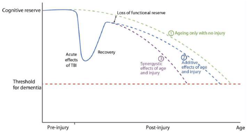
Any injury to the brain can generate significant loss of brain volume, which in turn can initiate neuropathological changes, such as the presence of certain biomarkers (β-amyloid and tau), as well as neuroinflammatory changes. These changes can cause a younger aged brain to have symptoms that resemble an older neurodegenerative brain.
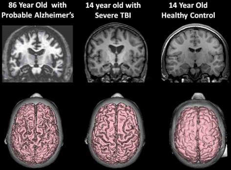
Using MRI scans and sophisticated software tools for quantitative imaging (NeuroQuant) to analyze the resulting images has become a significant element in the assessment of patients with TBI. Volumetric MRI studies performed after a brain injury can provide physicians with regional and whole brain volumes, which can be followed over time. This is especially meaningful, as global and regional brain atrophy from a single minor brain injury or concussion, can be detected one year after the injury.
NeuroQuant is a leading medical device software that automatically and accurately segments and calculates brain structure volumes from the 3D MR images, thereby providing anatomic information about the brain in a non-invasive fashion.
For physicians evaluating TBI, the Triage Brain Atrophy report provides volume measurements of 39 left and right brain structures and indicates the patient’s brain structure volume measurements as compared to a healthy population (Cortechs.ai’ normative reference data). This information provides physicians with an accurate indication of where an individual’s brain structure volumes are compared with their age- and gender-matched peers. The Triage Brain Atrophy report provides critical supporting quantitative volumetric data to aid physicians in their assessment of TBI.
References:
[1] Bigler, Erin D. (2013). Traumatic brain injury, neuroimaging and neurodegeneration. Frontiers in Human Neuroscience, 7(395):1-15.
[2] Zhou, Y., Kierans, A., et al. (2013). Mild traumatic brain injury: longitudinal regional brain volume changes. Radiology: (3) 267:880-890.