As the population ages, the demand for early and accurate assessment of neurodegenerative diseases continues to grow. Clinicians are increasingly asked to distinguish between normal aging and early dementia, which is a distinction that can be subtle on conventional MRI.
Traditional visual interpretation, while essential, often struggles to capture small but clinically meaningful changes in brain volume over time. This is where quantitative imaging provides a new layer of insight.
Why Quantitative MRI Matters
Visual reads of MRI scans can vary between readers and across time, particularly when atrophy occurs gradually. Quantitative volumetric analysis bridges this gap by providing objective, reproducible measurements that complement clinical evaluation.
Recognizing Disease-Specific Atrophic Patterns
Beyond generalized volumetric analysis, the NeuroQuant® Dementia: Age-Related Atrophy report helps clinicians recognize region-specific atrophy patterns associated with various neurodegenerative disorders.
Volumetric Data of Key Structures
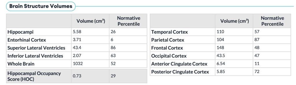
The NeuroQuant Dementia: Age-Related Atrophy report transforms standard MRI data into precise, objective measurements that support the detection and monitoring of neurodegeneration. The NeuroQuant Dementia: Age-Related Atrophy report presents volumetric information and normative database comparisons of key structures relevant to dementia, such as hippocampi, entorhinal cortex, superior and inferior lateral ventricles, lobar cortices, and anterior and posterior cingulate cortices.
Longitudinal Assessment
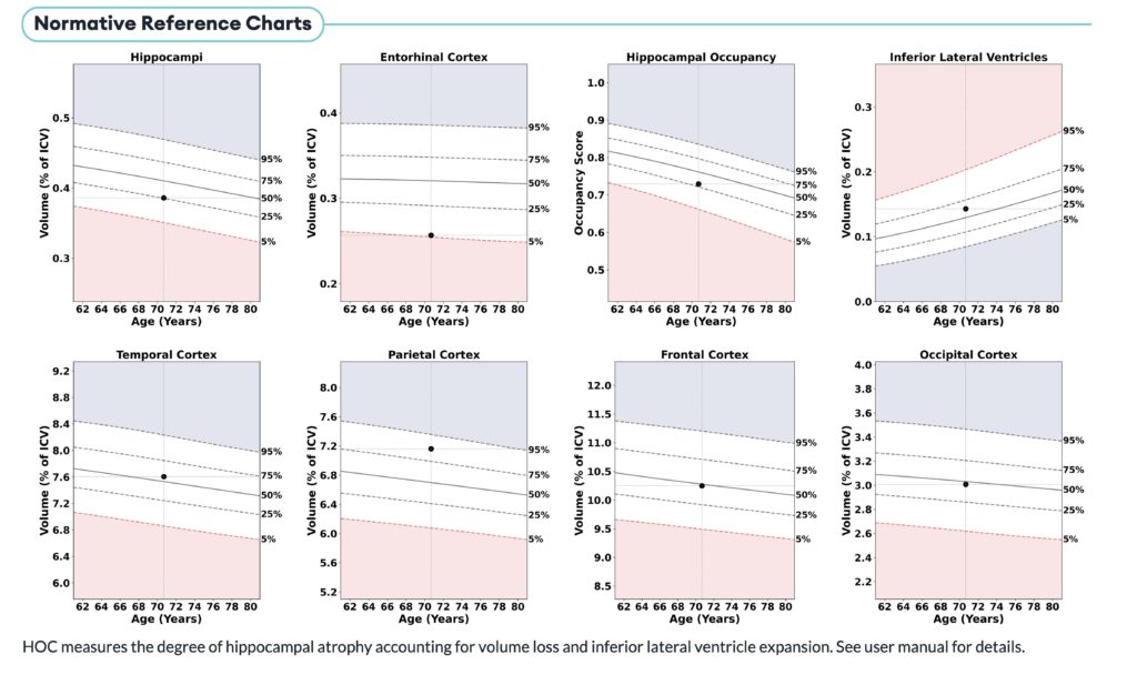
In addition to providing structural volumes of key structures, the NeuroQuant Dementia: Age-Related Atrophy report provides longitudinal comparisons to prior studies to help assess disease trajectory over time. This information is artfully displayed in Normative Reference Charts in the report, providing a visual representation of disease surveillance compared to age and sex-matched norms.
Most Common Types of Dementia
Dementia encompasses multiple neurodegenerative and vascular conditions, each with distinct imaging and clinical features. Understanding these patterns helps clinicians interpret quantitative MRI results effectively and identify disease-specific atrophy signatures. The NeuroQuant Dementia: Age-Related Atrophy report supports clinicians in their assessment of atrophic patterns that are characteristic of the most common types of dementia:
1. Alzheimer’s Disease (AD)
- Prevalence: 60–70% of all dementia cases.
- Pathology: Beta-amyloid plaques and tau tangles causing neuronal loss, particularly in the hippocampus and medial temporal lobes.
- Symptoms: Progressive memory loss, impaired language, visuospatial deficits.
- MRI Pattern: Hippocampal and medial temporal atrophy with ventricular enlargement.
- NeuroQuant Marker: Low hippocampal volume with compensatory expansion of the inferior lateral ventricles. This pattern is typical of AD, and is further characterized by the Hippocampal Occupancy (HOC) Score on the NeuroQuant Dementia report.
The HOC score is an arithmetic mean of ratios of hippocampal volumes to the sum of the hippocampal and inferior lateral ventricle (ILV) volumes in each hemisphere separately. It is an estimate of medial temporal lobe atrophy based on inferior lateral ventricle expansion and hippocampal atrophy. Given this ratio of hippocampal volume to ILV volume, a low HOC score can be associated with an increased likelihood of AD.
2. Lewy Body Dementia (LBD / DLB)
- Prevalence: 10–15% of cases.
- Pathology: Alpha-synuclein (Lewy body) deposition in cortical and brainstem neurons.
- Symptoms: Fluctuating cognition, visual hallucinations, REM sleep disturbance, parkinsonism.
- MRI Pattern: Posterior cortical and occipital atrophy with relative hippocampal preservation.
- NeuroQuant Marker: Posterior cortical gray matter reduction without significant hippocampal atrophy.
LBD typically manifests as occipital lobe and posterior parietal volume loss on MRI. The NeuroQuant Dementia: Age-Related Atrophy report can help point to this distinctive pattern by displaying cortical atrophy in the occipital and posterior parietal regions with preservation of frontal and temporal lobe cortices and subcortical structures.
4. Frontotemporal Dementia (FTD)
- Prevalence: 5–10%, especially in patients under 65.
- Pathology: Tau or TDP-43–related degeneration of frontal and anterior temporal lobes.
- Symptoms: Personality change, disinhibition, or language impairment.
- MRI Pattern: Frontal and temporal lobe atrophy, often asymmetric. Relative preservation of hippocampi.
- NeuroQuant Marker: Reduced frontal and temporal gray matter volumes
FTD patterns are consistent with frontal and anterior temporal lobe atrophy with preserved hippocampal/medial temporal lobe volumes. This characteristic neurodegeneration can easily be appreciated on the NeuroQuant Dementia: Age-Related Atrophy report.
By quantifying these distinct patterns over time, NeuroQuant helps clinicians correlate imaging findings with cognitive and behavioral symptoms, supporting more accurate disease tracking and management. Recognizing these characteristic atrophy patterns enhances diagnostic accuracy and helps clinicians use NeuroQuant data to distinguish between normal aging and specific disease states.
Clinical Value Across the Care Team
- Neurologists: Gain objective data to support diagnosis and treatment planning.
- Radiologists: Add quantitative precision to dementia imaging reports.
- Researchers: Standardize volumetric endpoints across multi-site studies.
- Patients: Visualize their brain health trajectory clearly over time.
Conclusion
Quantitative MRI with NeuroQuant transforms brain imaging into a measurable biomarker for neurodegeneration. By combining volumetric data with clinical expertise, clinicians can track subtle changes, differentiate disease states, and guide personalized care for patients experiencing cognitive decline.