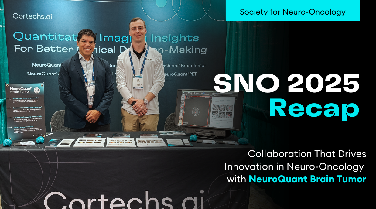
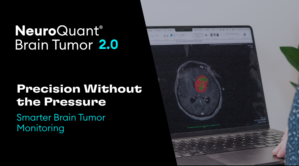
- NeuroQuant® Brain Tumor
FDA-cleared software for advanced brain tumor analyses
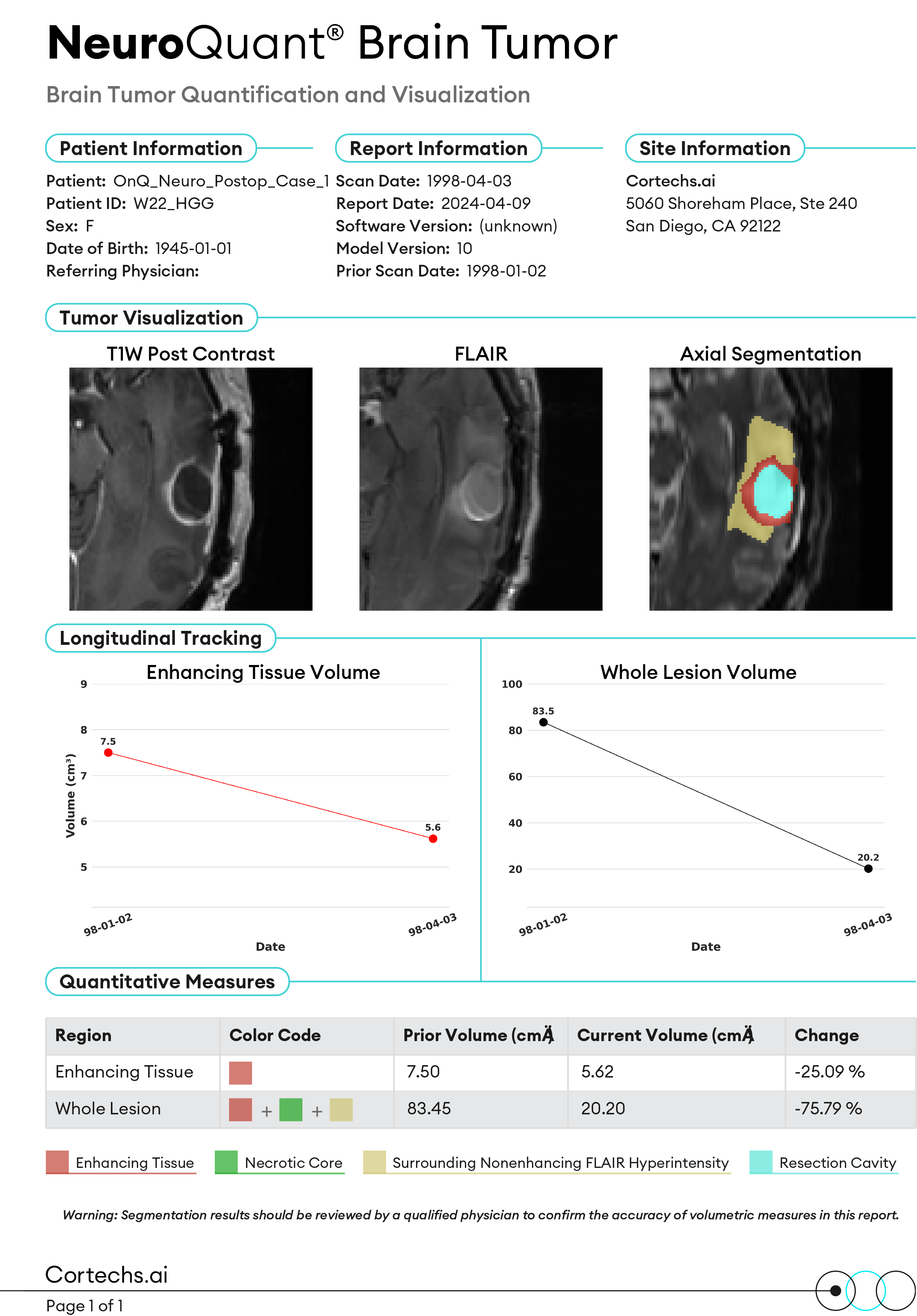
Powered by Al, NeuroQuant® Brain Tumor assists radiologists, oncologists, neurosurgeons, and the Tumor Board by providing objective quantification and analysis of tumor changes over time
Comprehensive Brain Tumor Analysis
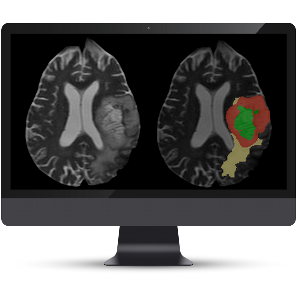
- Glioma Segmentation
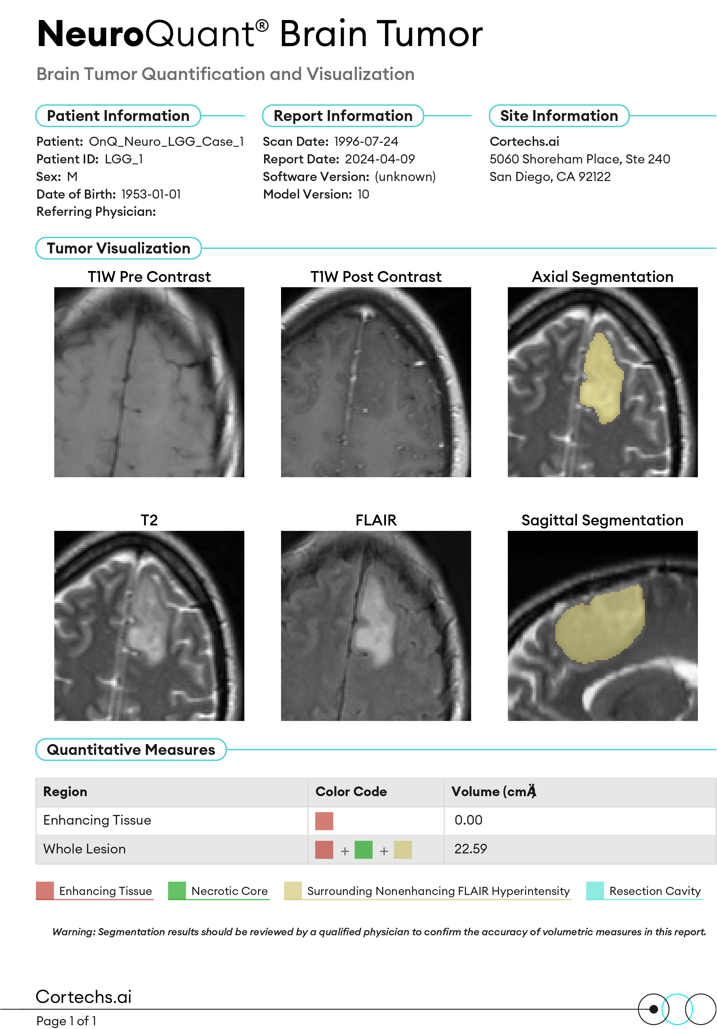
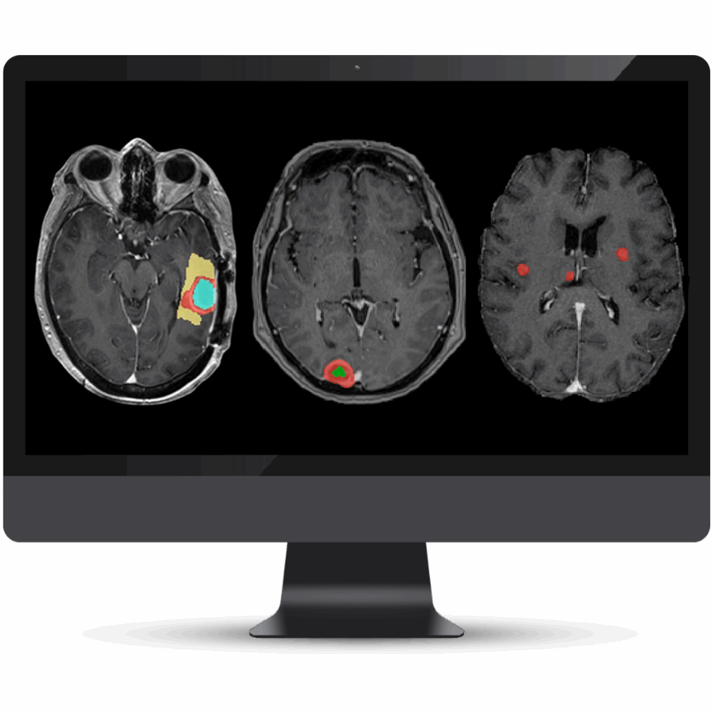
Advanced classification and segmentation of both high-grade and low-grade gliomas in pre- and postoperative studies. Our AI analyzes regions of interest including the necrotic core, enhancing tumor tissue, surrounding FLAIR hyperintensity, and resection cavity.
Required sequences: T1 pre, T1 post, T2, and T2 FLAIR
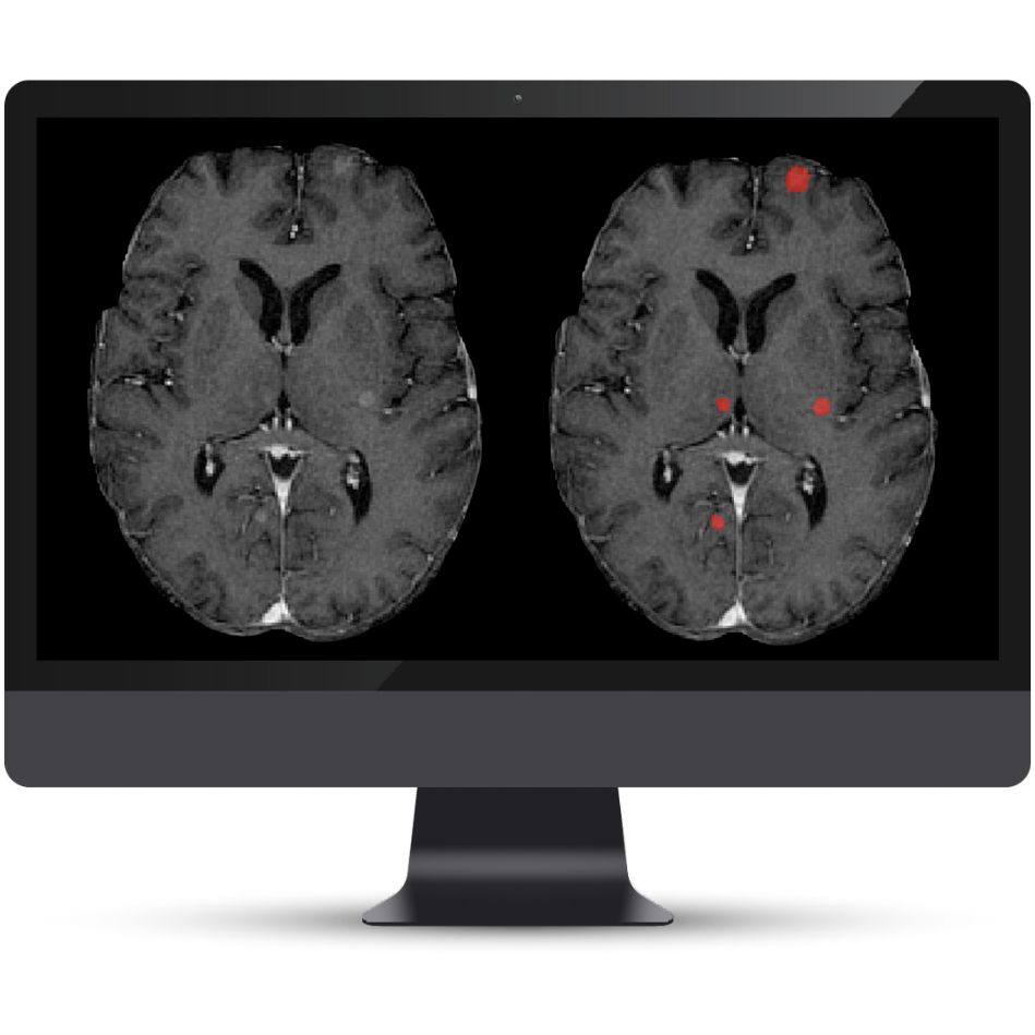
- Metastasis Segmentation
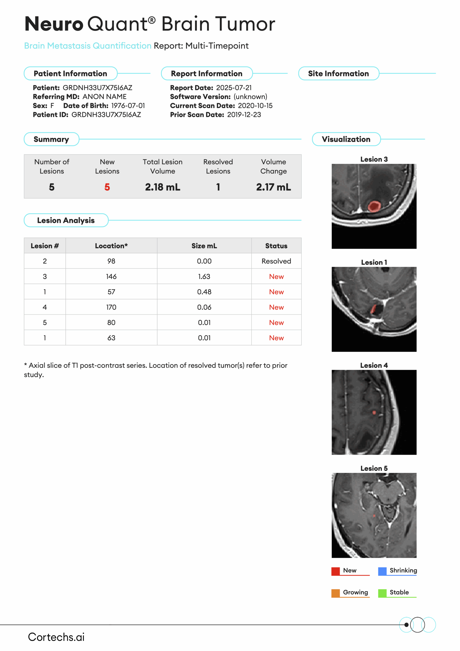
Designed for both single- and multi-timepoint studies, this module classifies and tracks brain metastases over time. It analyzes new, enlarging, shrinking, stable, and resolved lesions with comprehensive output including lesion count, individual and total volumetrics, and precise lesion location.
Required sequences: T1 pre and T1 post
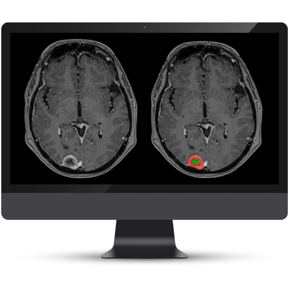
- Meningioma Segmentation
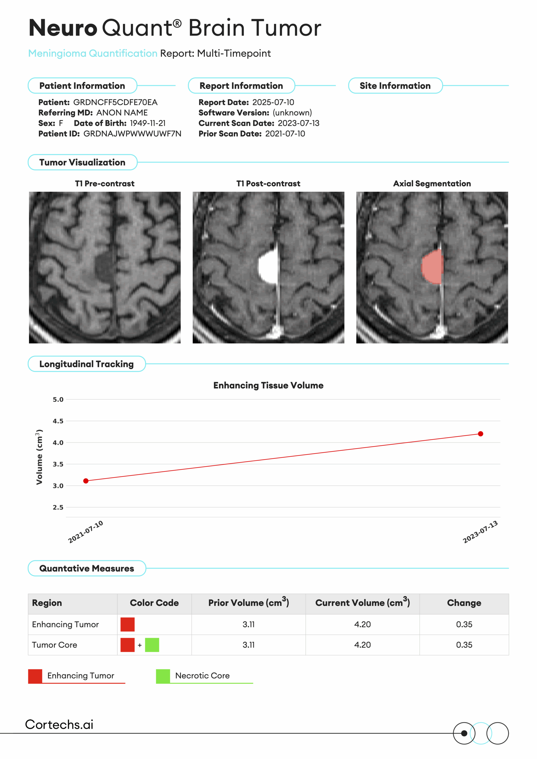
Supports both single- and multi-timepoint analysis of meningiomas. The system segments the necrotic core and enhancing tissue, providing detailed volumetric data for current, prior, and interval change analysis.
Required sequences: T1 pre and T1 post
Objective Tumor Insights Made Simple

Segmentation & Volumetrics Report
Detailed volumes of tumor regions are provided in comprehensive reports and automatically routed to PACS for seamless integration into your workflow.

Longitudinal Analysis
Prior studies of the same patients are automatically included on reports for precise volumetric comparison over time, enabling better treatment monitoring.
Tumor Segmentation Color Overlay Series
Slice-by-slice tumor segmentations are routed to PACS with tumor regions of interest color coded for easy viewing and interpretation.
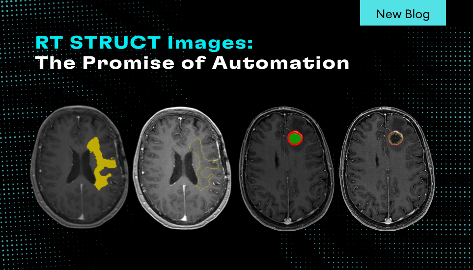
RT STRUCT
Advanced contour generation that can be manipulated for treatment planning, surgical planning, and radiation oncology applications.
NeuroQuant® Brain Tumor Benefits
Enhanced Objectivity & Reproducibility
Quantitative brain tumor volumetrics provide more objectivity and are more reproducible than 2D visual analyses ¹
¹ Blomstergen et al., (2018, July)
Time-Series Analysis
Displays time-series data for tumor volumes across multiple scans, allowing for better comparison and treatment planning
Comprehensive Tumor Coverage
Supports the analysis of gliomas, metastases, and meningiomas, offering broader clinical utility than solutions limited to one tumor type
NeuroQuant® Brain Tumor Reports
Comprehensive segmentation and volumetrics reporting, automatically routed to PACS.

Understanding CPT Codes 0865T & 0866T
As of January 1, 2024, new Category III CPT codes (0865T, 0866T) are active for AI-assisted quantitative brain MRI analysis. These codes apply to a variety of vendor solutions, including Cortechs.ai, and serve as an essential step toward achieving permanent reimbursement.
