By
Cortechs.ai
2 mins
In case you haven’t heard the news, LesionQuant is now available!
On August 3, we released LesionQuant™, a notable enhancement to our trusted and proven MRI volumetric image analysis solution, NeuroQuant®. LesionQuant provides fast, accurate and automated FLAIR lesion and brain volume measurements, as well as, lesion visualization.
LesionQuant combines 2D or 3D FLAIR with 3D T1 MRI images to quantify and visualize FLAIR lesions and brain structure volumes. When using LesionQuant, physicians have access to additional supportive volumetric data that can enrich their clinical treatment planning and disease progression monitoring of patients with white matter diseases, such as Multiple Sclerosis.
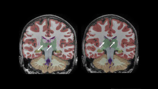
LesionQuant’s FLAIR lesion overlay provides two different DICOM files for review: FLAIR lesion segmentation and FLAIR lesion change, for physicians to easily review color-coded lesion segmentation, slice-by-slice, on a PACS or other DICOM viewer. The video below provides an example demonstration of sample slices that can be viewed of LesionQuant’s FLAIR lesion overlay.
FLAIR lesion segmentation:
Lesions are color-coded* based on where they are anatomically located in the brain.
| Leukocortical (Red) |
Periventricular White Matter (Purple) | Infratentorial (Orange) |
Deep White Matter (Yellow) |
FLAIR lesion change:
Lesions are color-coded to distinguish between increasing and decreasing volumes.
(when current and prior scans are submitted)
Interested in learning more about LesionQuant?
We will be hosting a webinar: An Introduction to LesionQuant on Tuesday, October 11 at 11 am PT. Sign up here.
Are you attending ECTRIMS 2016?
Stop by Stand D08 at ECTRIMS in London next week and let us provide you with additional information. In the meantime, contact us with questions, or to set up a demonstration/meeting at ECTRIMS.
Request 5 free trial scans of LesionQuant now.
![]()
* Colors may appear lighter or darker due to image intensities.
![]()
Share