In 1996 FreeSurfer§, and later NeuroQuant® in 2006, revolutionized image segmentation by introducing software for the automation of processing and analyzing MRIs. The Bayesian approach used for this automatic image segmentation was based on a static atlas, which works very well for a large part of the population but shows limitations when the subject’s anatomy is too far from the generic norm or too far outside the standard cohort age range.
One June 1, 2015, Cortechs.ai released the patented Dynamic Atlas™ developed for NeuroQuant 2.0, which introduces a new unique, approach allowing automatic segmentation and volume measurements for patients ranging from 3-100 years, and furthering robustness of segmentation and goodness-of-fit across all age ranges to atlas.
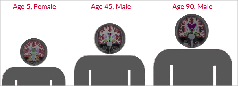
The Dynamic Atlas creates a personalized atlas instantaneously based on patient specific covariates and prior anatomical knowledge derived from a large database. The prior knowledge is represented by the target image, likelihoods, and class priors, which are compiled over a large cohort of individuals.
Using proprietary algorithms, the patients observed data, which is comprised of original image data and derived data from image registration and segmentation, is further processed to predict the atlas parameters for an individual. An individual’s atlas parameters then form the basis of the personalized atlas allowing for the segmentation of virtually any patient instantly.
Want to learn more about Cortechs.ai Dynamic Atlas? Cortechs.ai has developed a Dynamic Atlas white paper, which is now available for download.
[§] The FreeSurfer software application for cortical reconstruction and volumetric segmentation was originally developed by Anders M. Dale et al. at Massachusetts General Hospital/Harvard Medical School and is jointly owned by Cortechs.ai and Massachusetts General Hospital.