Cortechs.ai | Diffusion-weighted imaging in cancer

Diffusion-weighted imaging in cancer

Diffusion-weighted imaging in cancer: physical foundations and applications of Restriction Spectrum Imaging

In this review, we provide a detailed explanation of the biophysical basis of diffusion contrast, emphasizing the difference between hindered and restricted diffusion, and elucidating how diffusion parameters in tissue are derived from the measurements via the diffusion model. We describe one advanced DWI modeling technique, called restriction spectrum imaging (RSI). 2014.

[button]Download[/button]

More Resources

08/18/2025

Why Bigger Normative Databases Mean Better Brain Imaging

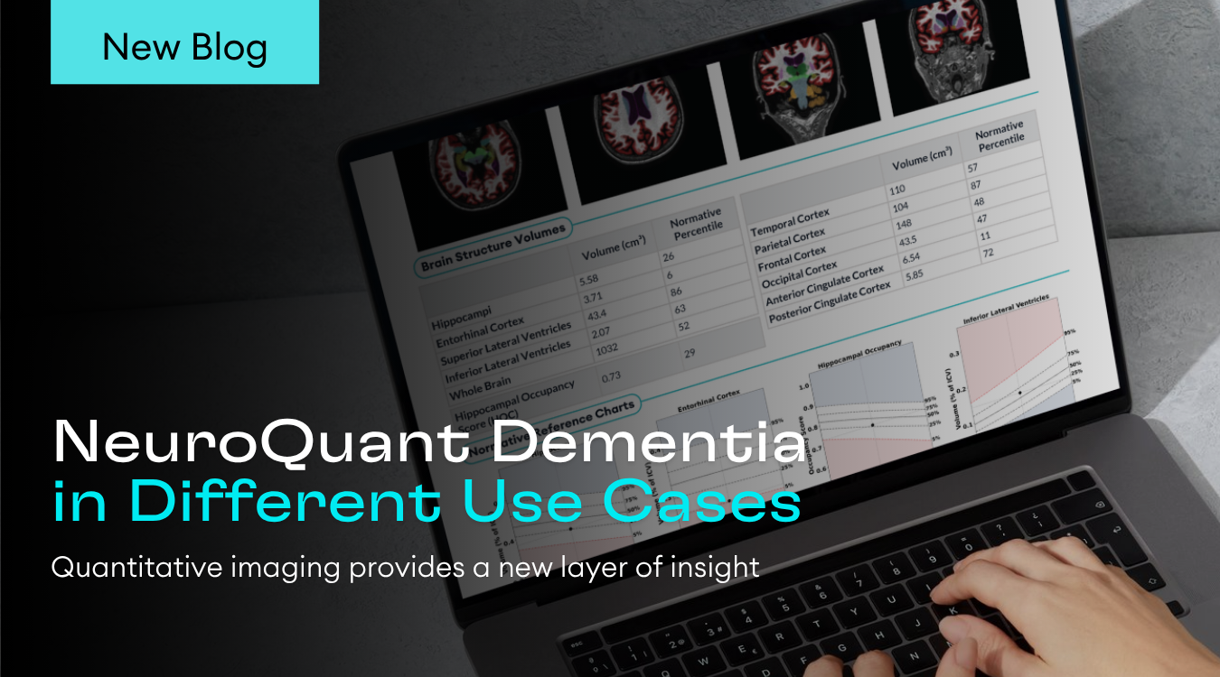
NeuroQuant® 5.2 compares patient brain MRI volumes to over 7,000 healthy scans, giving clinicians clear, percentile-based context for better assessments.

08/07/2025

Navigating Reimbursement for NeuroQuant: Understanding CPT Codes 0865T & 0866T 

Let’s break down the latest category III CPT codes and example clinical use cases that illustrate their value.

07/31/2025

Precision Without the Pressure: Smarter Brain Tumor Monitoring

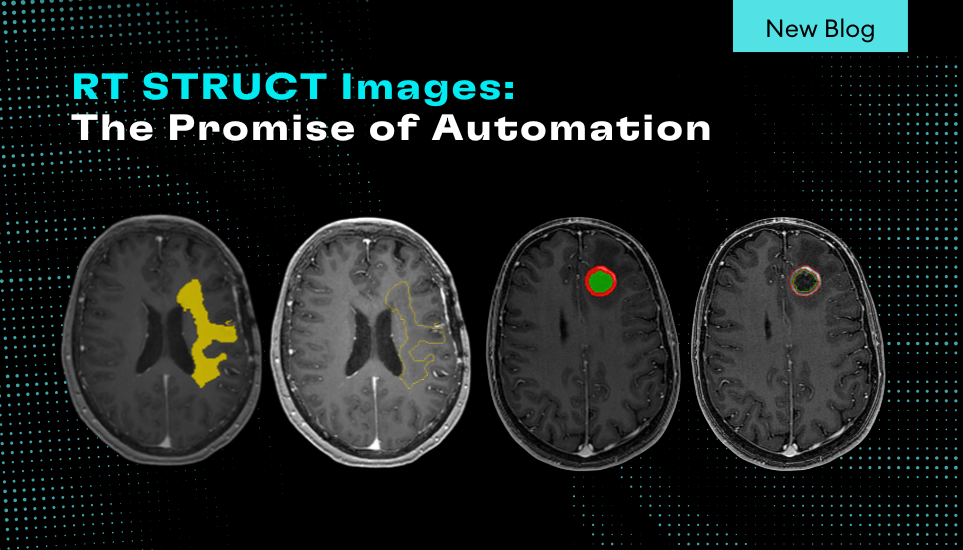
Learn about NeuroQuant Brain Tumor 2.0 and how it assists clinicians by providing objective quantification and analysis of tumor changes over time.

07/30/2025

Cortechs.ai Announce Next-Gen NeuroQuant Brain Tumor: AI-Driven Metastasis & Meningioma Segmentation 

NeuroQuant Brain Tumor is the first FDA-cleared, cloud-native tool to offer automated volumetric segmentation for brain metastases, meningiomas, and gliomas.

07/28/2025

Longitudinal Assessment of Traumatic Brain Injury Using NeuroQuant®

A case study with Dr. Suzie Bash on NeuroQuant as a reliable, reimbursable, and efficient tool in the evaluation and follow-up of TBI.

07/24/2025

Understanding the Hippocampal Occupancy Score

One of the most frequently asked questions among customers is, “What does the Hippocampal Occupancy Score (HOC) represent within the Dementia Report?”
Scroll to Top