Automated volumetric MR imaging provides an opportunity to utilize brain structure volume measurements to assess symptoms of post-traumatic stress disorder (PTSD) in patients by analyzing the volumes of affected brain regions correlated with PTSD, tracking and evaluating them over time. PTSD can develop in people experiencing or witnessing a life-threatening event, like combat, a natural disaster, a car accident, or sexual assault.
PTSD researchers are currently focusing on volume loss in the amygdala, the ventromedial prefrontal cortex and the hippocampus, which may be affected in response to trauma and chronic stress. The Triage Brain Atrophy report provides the left, right and total normative percentile for the amygdalae, hippocampi and the medial orbitofrontal (considered anatomically synonymous with the ventromedial prefrontal cortex), as well as an overview of 41 additional structures/regions, including the whole brain volume.
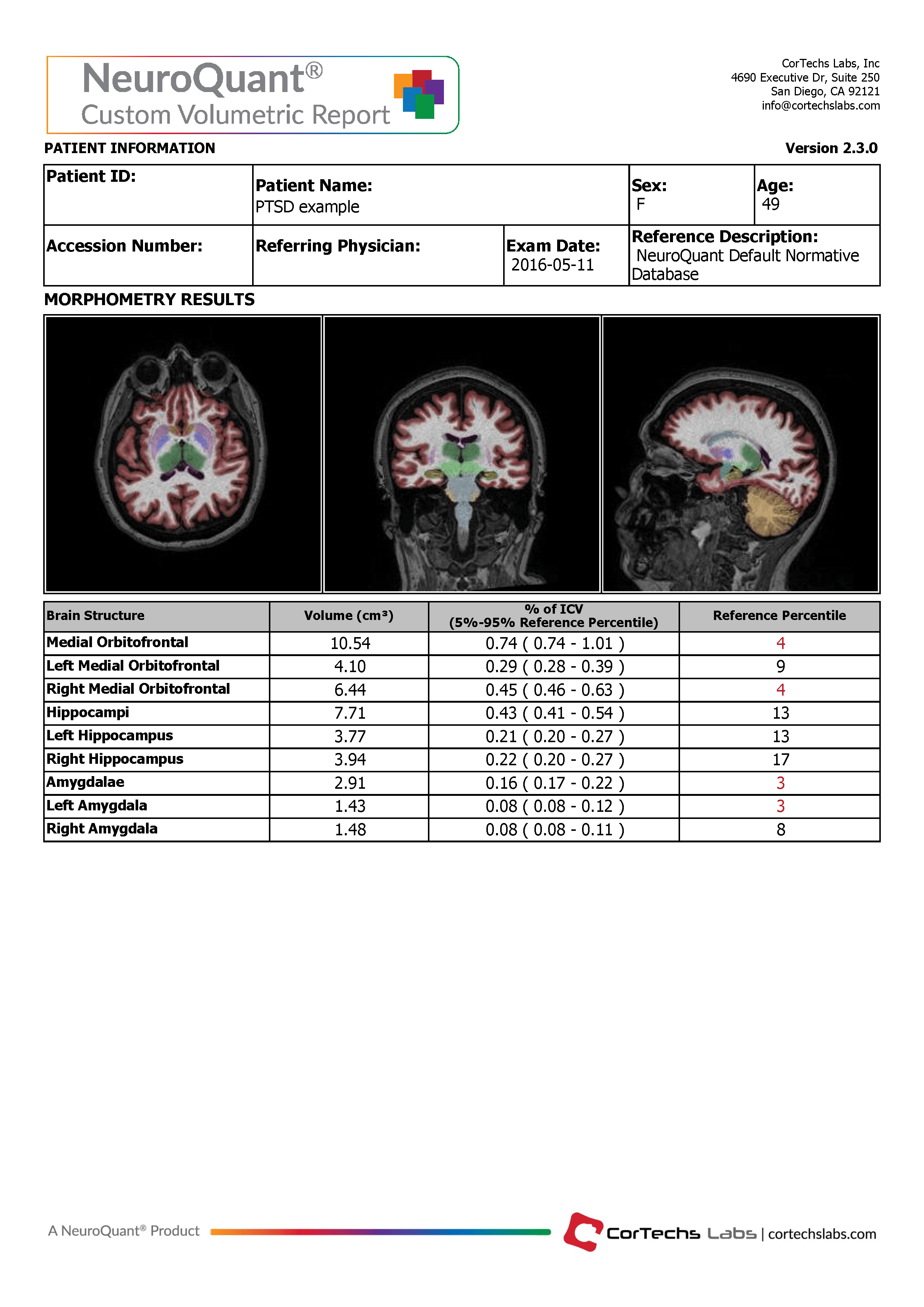
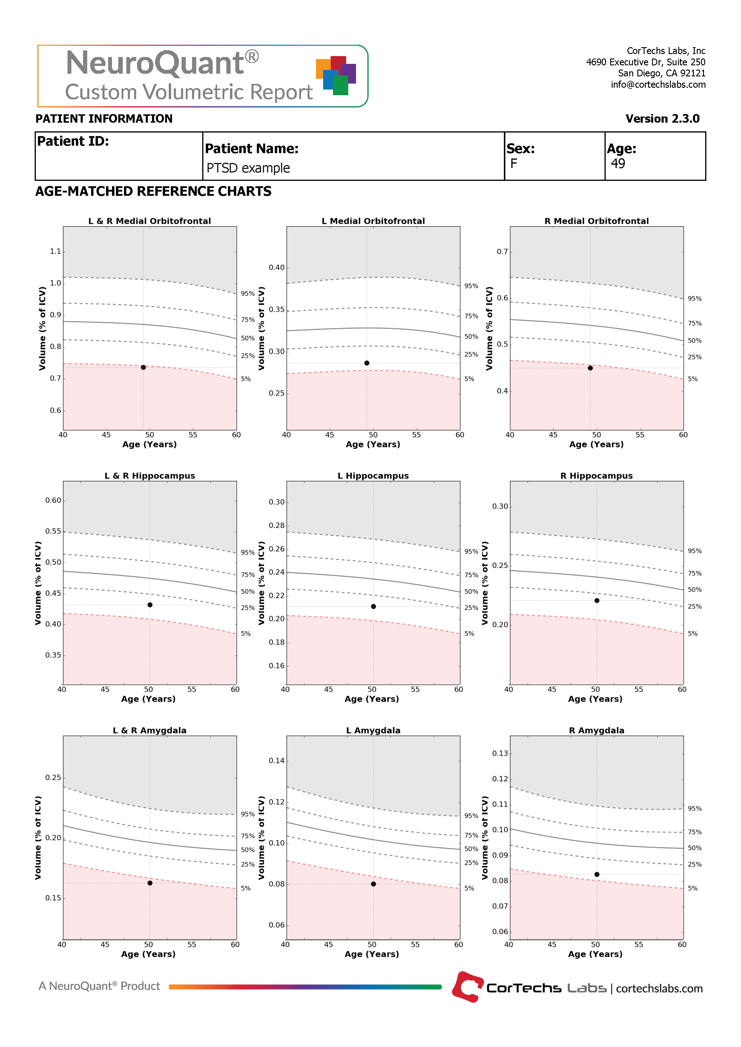
For clinicians with a focus on assessing PTSD patients, the Custom Volumetric report can provide brain structure volume measurements for the amygdala, the ventromedial medial prefrontal cortex and the hippocampus. These structures can be divided by right, left, total and the asymmetry between the right and left hemispheres. The resulting volumes are compared to a healthy population via Cortechs.ai’ normative reference data. This allows NeuroQuant to deliver a precise indication of where that individual’s brain structure volumes lie within an age- and gender-based reference chart.
Since PTSD can happen at any age, NeuroQuant reports are applicable for ages 3 to 100 years. Additionally, the clinician is able to review axial, coronal and sagittal DICOM images in which the segmented brain are given a color overlay.
The NeuroQuant Custom Volumetric report provides a multi time point feature, which can be used to assess volume changes in these brain structures by displaying the current measurement together with all previous measurements on the normative reference charts. Physicians can use this information to quickly and easily evaluate longitudinal change in several cortical and subcortical structures.
Lopez, Katherine C. et al. (2017) “Brain Volume, Connectivity, and Neuropsychological Performance in Mild Traumatic Brain Injury: The Impact of Post-Traumatic Stress Disorder Symptoms.” Journal of Neurotrauma 34.1: 16–22.
Morey RA, Gold AL, LaBar KS, et al. (2012) Amygdala Volume Changes in Posttraumatic Stress Disorder in a Large Case-Controlled Veterans Group. Arch Gen Psychiatry. 69(11):1169–1178.
