Microvascular ischemic disease (MVID) has emerged as a growing concern as the long-term consequences of its progression are increasingly recognized. Its association with cognitive decline, stroke, and dementia has made MVID a key focus in neurological research and clinical management. This heightened attention underscores the need for early detection, standardized diagnosis, and quantitative assessment. The NeuroQuant Microvascular Report delivers valuable insights into the imaging features of microvascular ischemic disease. By providing precise, reproducible quantification of white matter hyperintensity burden, regional distribution, and automated Fazekas scoring, it offers objective data to support clinical evaluation and informed decision-making.
What is Microvascular Ischemic Disease?
Microvascular ischemic disease (MVID), or cerebral small vessel disease (SVD), is the most common cerebrovascular disease.1 It is a condition characterized by the blockage of small vessels in the brain. Chronic blockages of tiny vessels, such as arterioles and capillaries, can result in ischemic brain changes due to reduced blood supply over time.
The risk factors for microvascular ischemic disease lie in parallel to many other disease states. Risk factors include high blood pressure, diabetes, obesity, smoking, and advanced age. Age is the most common risk factor, making microvascular ischemic disease of particular concern for the geriatric population. The presence of microvascular ischemic disease increases with age, with a prevalence of over 90% after age 75.2
Microvascular ischemic disease, when risk factors are uncontrolled, may lead to cognitive decline and chronic vascular brain injury such as vascular dementia or ischemic stroke. Vascular dementia is the second most common form of dementia after Alzheimer’s disease and constitutes 15-20% of all dementia cases. 1 The risk for ischemic stroke also increases with microvascular ischemic disease. Studies have shown that microvascular ischemic disease may cause 25% of strokes.
Imaging Characteristics of Microvascular Ischemic Disease
The primary imaging characteristic of microvascular ischemic disease is white matter hyperintensities (WMH). WMH appear as bright (hyperintense) lesions on T2 and T2 FLAIR MRI imaging. In small vessel disease, lesions are often present in the periventricular and deep white matter regions (supratentorial regions).3 While rarer and indicative of alternate disease presentation, WMH can also exist in the cerebellum and brainstem (infratentorial region). Radiographic interpretation of WMH on MRI may include Fazekas scoring, which is a visual grading tool to characterize the deep white matter and periventricular WMH burden.
Since white matter lesions can exist in multiple vascular or inflammatory conditions, differential diagnosis between MVID and other pathophysiologic conditions exhibiting white matter lesions can be challenging and is often dependent on patient presentation, demographics, and symptomology.
STRIVE: An Effort to Streamline Reporting
In 2013, a standardization in the imaging assessment of small vessel disease (SVD) was introduced. STRIVE (Standards for reporting of vascular changes in neuroimaging) guidelines were published in The Lancet. Establishing STRIVE guidelines was an international attempt aimed at categorizing small vessel disease imaging characteristics on a universal reporting scale. STRIVE aimed to achieve the following:, “first, to provide a common advisory about terms and definitions for features visible on MRI; second, to suggest minimum standards for image acquisition and analysis; third, to agree on standards for scientific reporting of changes related to SVD on neuroimaging; and fourth, to review emerging imaging methods for detection and quantification of preclinical manifestations of SVD. Our findings and recommendations apply to research studies, and can be used in the clinical setting to standardize image interpretation, acquisition, and reporting.” 3
In 2023, updates to STRIVE standards were released, termed STRIVE-2. While STRIVE-1 focused mainly on qualitative neuroimaging biomarkers, STRIVE-2 emphasized the importance of including quantitative measures, volumetric data, regional lesion distribution, and longitudinal tracking.4 The NeuroQuant Microvascular report functions to support STRIVE-2 guidelines and ease the reporting burden on radiologists.
How does the NeuroQuant Microvascular report assess small vessel disease?
NeuroQuant is an AI tool that provides quantitative brain volume information from MRI. The NeuroQuant Microvascular report utilizes 2 MRI sequences to aid in the assessment of SVD:
- 3D T1 GRE: NeuroQuant software performs volumetric segmentation of 3D T1 GRE and lists structures on the NeuroQuant Microvascular report that support assessment of disease state atrophic or enlargement patterns.
- 2D or 3D T2 FLAIR: NeuroQuant software analyzes WMH from T2 FLAIR MRI. Information on WMH volume, regional location, and Fazekas score are included in the NeuroQuant Microvascular report.
This multi-sequence assessment aids in WMH lesion and brain structure volume quantification. In addition to providing objective data, NeuroQuant Microvascular reports provide the strong clinical advantage of longitudinally tracking unlimited MRI time points to aid in disease surveillance.
Traditional Challenges of MVID Imaging Analysis
1. Analysis is subjective
In assessing microvascular ischemic disease imaging characteristics, no two radiologists will read an exam in the same way. Inter-reader variability can pose a challenge to neurologists in interpreting radiology reports and determining care pathways.
2. Standard radiology reads can lack precision
With an absence of quantitative volumetric information, radiology reporting tends to take a qualitative approach of describing WMH burden as “mild”, “moderate”, or “severe”. Traditional reads may overlook subtle disease progression.
3. Lack of standardized form of reporting
Although standards have been established on MVID/SVD reporting, lack of consistency still exists in many scenarios.
4. Small lesion changes are difficult to quantify over time
Accurate quantification of minute changes is very difficult to assess without volumetric measurement.
Clinical Benefit of the NeuroQuant Microvascular Report
1. Objective reporting
The NeuroQuant Microvascular report produces objective and reproducible quantification of WMH burden, as well as brain structure volumes and normative percentiles. Providing measurable data helps alleviate the inherently subjective nature of visual WMH analysis. Correlation of the NeuroQuant Microvascular report with patient presentation helps improve referring providers’ diagnostic confidence.
2. Precise measurements with automated Fazekas scoring
White matter hyperintensity burden is easily assessed with the NeuroQuant Microvascular report. The following measurements are included:
- FLAIR WMH Burden in mL
- FLAIR WMH regional location (supratentorial or infratentorial)
- Fazekas score of 0-3
- 0 corresponds to ≤ 1.5mL (none or minimal FLAIR WMH)
- 1 corresponds to > 1.5mL (multiple punctuate FLAIR WMH)
- 2 corresponds to > 10mL-35mL (beginning confluency of FLAIR WMH)
- 3 corresponds to > 35mL (large confluent FLAIR WMH)
- Brain structure volumes susceptible to volume changes with increased WMH burden
- Whole brain
- Cortical gray matter
- Cerebral white matter
- Ventricles
- Cortices (temporal, parietal, frontal, occipital)
- Brain structure volume normative percentiles
3. Standardized format
The NeuroQuant Microvascular report gives comprehensive data presented in the same format across patients and time points, providing simplified and streamlined access to pertinent values. Analysis of WMH volumes, WMH regional distribution, Fazekas score, brain structure volumes, and brain structure normative percentiles all support standardization in accordance with the STRIVE-2 criteria.
4. Longitudinal tracking
Comparison of prior and follow-up studies is an important tool in assessing patient disease progression. In the NeuroQuant Microvascular report, longitudinal analysis of WMH burden volume, WMH regional distribution, Fazekas score, and brain structure volumes are tracked and measured across time points to aid in disease surveillance.
Case Example
Clinical Background
- Patient: 85-year-old female
- Medical History: Memory loss, obesity, hypertension
- Symptoms: Presented for MRI with chief complaint of cognitive decline and gait disturbance
MRI Analysis
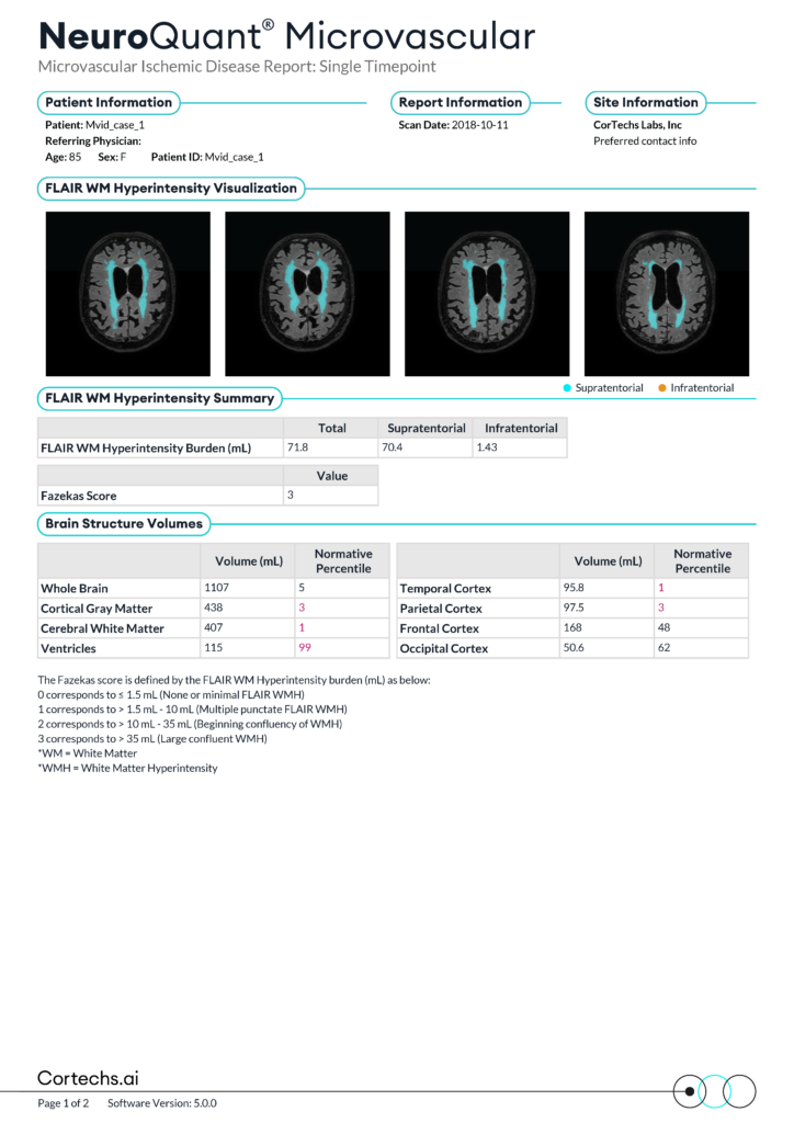
A standard brain MRI was performed on the patient, including 3D T1 GRE and T2 FLAIR sequences. The images were submitted for NeuroQuant Microvascular analysis.
Findings
- FLAIR WMH Burden Total: 71.8mL
- Supratentorial: 70.4mL
- Infratentorial: 1.43mL
- Fazekas score: 3 (large confluency of white matter)
- Brain structure volumes:
- Whole brain: 5th normative percentile for age and sex
- Cortical gray matter: 3rd normative percentile for age and sex
- Cerebral white matter: 1st normative percentile for age and sex
- Ventricles: 99th normative percentile for age and sex
- Temporal cortex: 1st normative percentile for age and sex
- Parietal cortex: 3rd normative percentile for age and sex
- Frontal cortex: 48th normative percentile for age and sex
- Occipital cortex: 62nd normative percentile for age and sex
Clinical Significance
The patient has a large portion of periventricular WMH, supporting their reported symptoms of cognitive decline. The patient has a small portion of infratentorial lesions, consistent with their complaint of gait disturbance
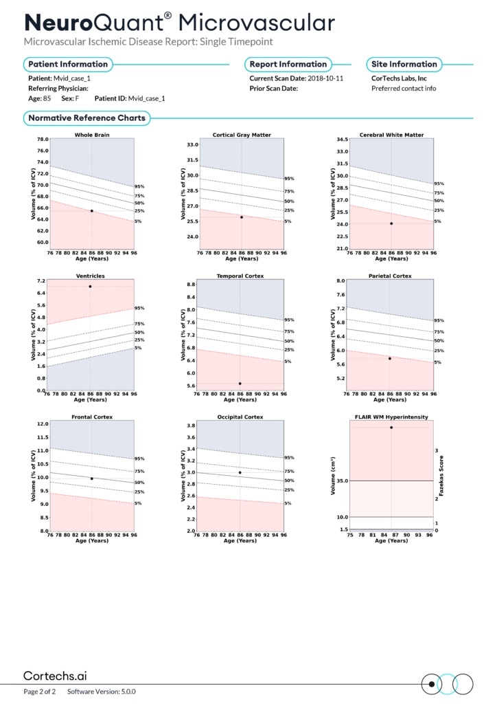
Statistically significant low volumes of the cortical gray matter, cerebral white matter, temporal and parietal cortex, and statistically significant high volume of the ventricles strongly supports a pattern of global atrophy consistent with small vessel disease burden. Low temporal lobe volume could point to a mixed presentation of vascular dementia and Alzheimer’s disease.
Conclusion
Microvascular ischemic disease is a common neurological condition that contributes to disease risk factors such as stroke and vascular dementia. Accurate, objective data is important in the effort to standardize reporting on vascular lesions and their associated pathologies.
NeuroQuant Microvascular reports provide a useful tool in the quantification and longitudinal assessment of microvascular ischemic disease. Quantitative measurements of white matter lesion and brain structure volumes provide enhanced clinical insight, reduce inter-reader variability, provide advanced visibility into disease progression, and increase clinical confidence among radiologists and referring providers.
References
- Singh A, Bonnell G, De Prey J, et al. Small-vessel disease in the brain. Am Heart J Plus. 2023;27:100277. Published 2023 Feb 18. doi:10.1016/j.ahjo.2023.100277
- Rodriguez L, Araujo AT, Vera DD, et al. Prevalence and imaging characteristics of cerebral small vessel disease in a Colombian population aged 40 years and older. Brain Communications. 2024;6(2). doi:https://doi.org/10.1093/braincomms/fcae057
- Wardlaw JM, Smith EE, Biessels GJ, et al. Neuroimaging standards for research into small vessel disease and its contribution to ageing and neurodegeneration. The Lancet Neurology. 2013;12(8):822-838. doi:https://doi.org/10.1016/s1474-4422(13)70124-8
- Duering M, Geert Jan Biessels, Brodtmann A, et al. Neuroimaging standards for research into small vessel disease—advances since 2013. Lancet Neurology. 2023;22(7):602-618. doi:https://doi.org/10.1016/s1474-4422(23)00131-x
