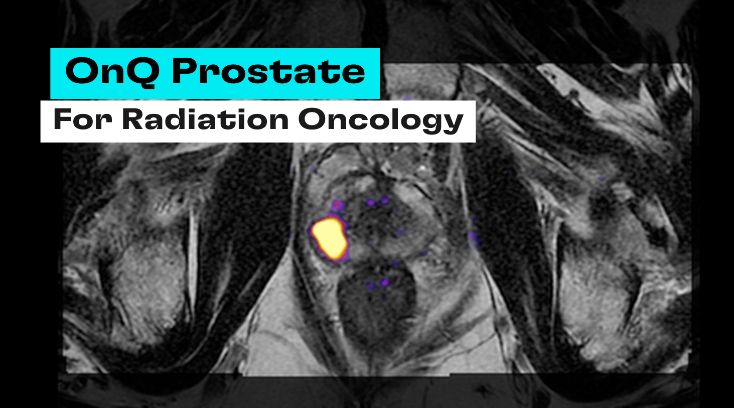
As the incidence of prostate cancer continues to rise globally, the need for advanced diagnostic tools has become increasingly urgent. In recent years MRI has been established as a cornerstone in the standard of care for prostate cancer detection, resulting in a rapidly growing number of prostate MRI exams and the corresponding demand for expert radiology interpretations. However, the inherent limitations of conventional mpMRI make it difficult to provide the level of accuracy and reproducibility required for optimal patient care. OnQ Prostate offers an FDA-cleared, state-of-the-art solution to address these challenges head-on.
OnQ Prostate employs Restriction Spectrum Imaging (RSI), an advanced diffusion-weighted MRI technique that was developed and patented by the University of California, San Diego, and licensed exclusively to Cortechs.ai. RSI has been identified as an emerging imaging biomarker because of its ability to hone in on intracellular restricted diffusion, a hallmark characteristic of aggressive cancer cells. As a result, the conspicuity of suspicious lesions on MRI is greatly increased, as well as the specificity from better tissue characterization, making it unlike anything on the market. Studies have shown that RSI’s biophysical approach improves the detection, invivo characterization, and localization of clinically significant prostate cancer, with stronger correlating to underlying histopathology at the voxel level.
With OnQ Prostate, radiologists are able to gain a new level of confidence in their reads and ability to differentiate clinically significant cancer from benign or normal prostate tissue. This helps reduce inter-reader variability, leveling the playing field amongst radiologists and elevating the performance of the whole group. Even for expert radiologists, OnQ Prostate delivers immense value by reducing ambiguity, especially in complex or borderline cases where standard imaging falls short. This plays a critical role in the effort to reduce false positives and unnecessary biopsies, while also reducing false negatives and missing cancers that need to be diagnosed and treated. OnQ Prostate seamlessly integrates into the radiology workflow without requiring a separate workstation or extra clicks, which is ideal for optimizing efficiency.
Now that we are in the era of MRI-guided procedures, there is an unprecedented need for referring physicians (e.g. urologists and radiation oncologists) to actually interact with the images themselves. This has resulted in an even higher dependence on radiologists to provide accurate and precise annotations to target lesions. Since OnQ Prostate images are much easier and more intuitive for the non-radiologist to understand, there is now opportunity for increased transparency between radiologists, referring physicians, and patients.
Adoption of advanced imaging technologies will empower more-informed clinical decision-making and ultimately lead to better patient outcomes. In this rapidly evolving field, OnQ Prostate stands out as a potentially transformative solution to elevate the standard of care throughout the entire prostate cancer care pathway, from diagnosis to management to treatment.


