According to the MS International Federation, multiple sclerosis (MS) is one of the most common diseases of the central nervous system, and more than 2,300,000 people worldwide are living with the diagnosis of MS. MS is defined as an immune-mediated response, causing inflammatory demyelination (damage to the myelin sheath). It is this damage that is the cause of the various symptoms associated with MS (including blurred vision, weak limbs, tingling sensations, unsteadiness and fatigue).
However, research has provided increased knowledge about the disease and has made it apparent that MS has two major contributors, inflammation and neurodegeneration.
With the recognition of neurodegeneration as a contributing factor in MS, the evaluation of brain volume loss in a variety of brain regions is increasingly important. Not only for initial diagnosis, but also for assessing disease progression and the identifying different disease stages, as well as confirming the types of MS. Studying the impact of the affected brain structures and measuring the extent of the atrophy has brought more knowledge to the MS community. Researchers now recognize that atrophy occurs even in the early stages of MS, and not only as previously assumed at the late stages.
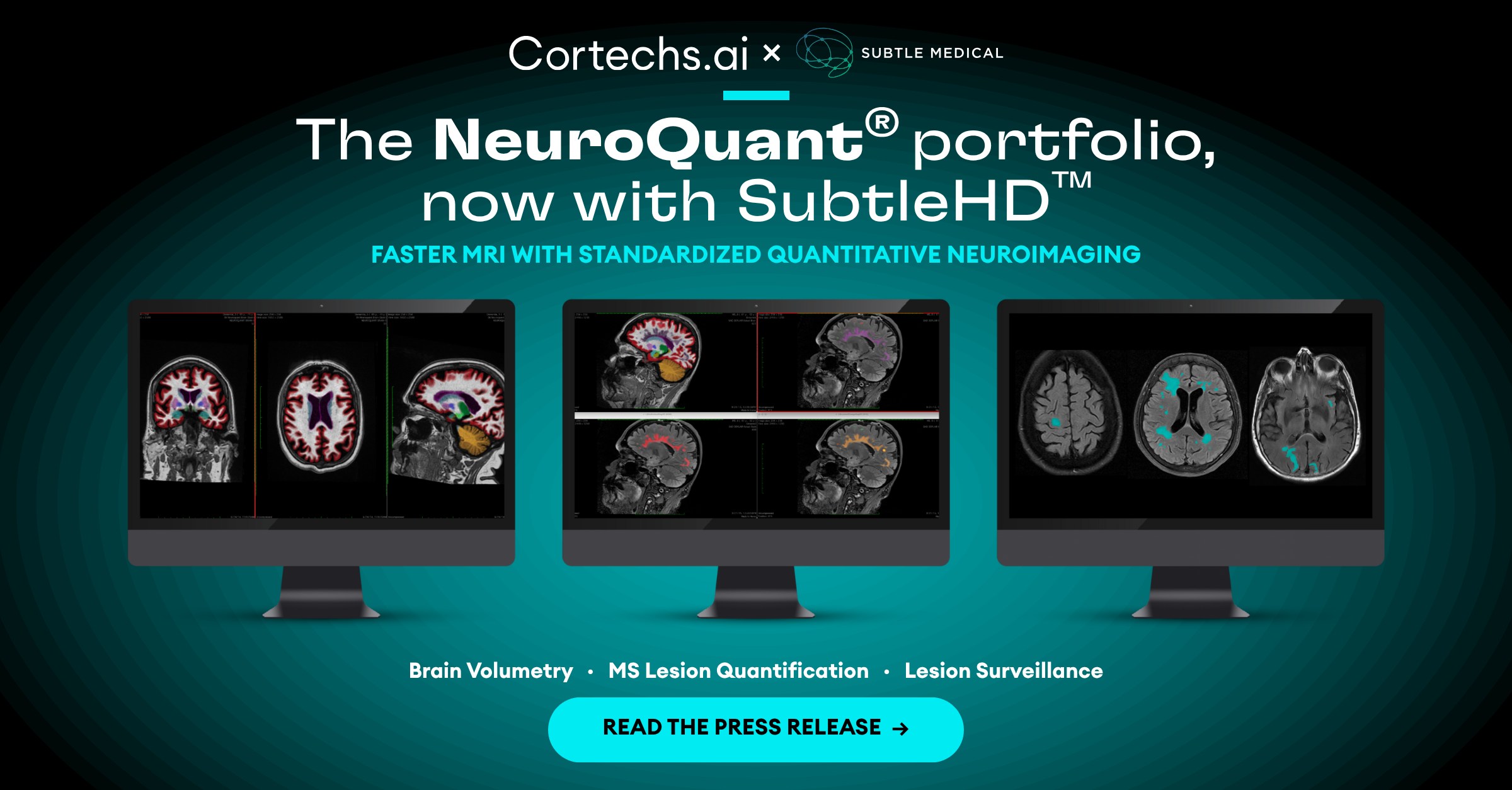
NeuroQuant® is a fully automatic, MRI post-processing solution that segments a large number of brain structures relevant to the assessment and ongoing monitoring of MS. It calculates the volumes and compares each volume to a normative database. This allows NeuroQuant to deliver a precise indication of where that individual’s brain structure volume lies within an age- and gender-based reference chart. The information provided by NeuroQuant allows a clinical user to take advantage of these research findings in evaluating their MS patients for the best possible care path.
- Raduea EW, Bendfeldta K, Mueller-Lenkea N, Magonc S, Sprengera T. Brain atrophy: an in-vivo measure of disease activity in multiple sclerosis. Swiss Med Wkly. 2013;143:w13887
- Barkhof F, Calabresi PA, Miller DH, Reingold SC. Imaging outcomes for neuroprotection and repair in multiple sclerosis trials. Nat Rev Neurol. 2009;5(5):256–66
- Compston A, Coles A. Multiple sclerosis. Lancet. 2002;359(9313):1221–31.