In the fast-paced realm of neurocritical care and neurosurgery, time-sensitive and precise diagnostic imaging can mean the difference between rapid intervention and devastating outcomes. Among the critical metrics derived from brain imaging, Lateral Ventricle Volume (LVV), Intracranial Volume (ICV), and Midline Shift (MLS) Index play pivotal roles in assessing and managing conditions like traumatic brain injury (TBI), stroke, hydrocephalus, and mass effect due to intracranial lesions. Thanks to advancements in imaging analytics and AI-driven automation, these volumetric and morphometric measures can now be rapidly quantified, offering clinicians a real-time window into intracranial pathology.
Critical Imaging Metrics
Lateral Ventricle Volume (LVV)
Changes in LVV can indicate a wide range of neurological issues, from hydrocephalus (ventricular enlargement) to ventricular compression due to mass effect. Quantifying LVV on CT offers:
- Objective markers for ventricular asymmetry
- Early detection of hydrocephalus or hemorrhagic ventricular compression
- Longitudinal monitoring in ICU patients
- Automated measurement enables reproducibility, which is essential in serial scans where subtle volumetric changes are clinically significant
Intracranial Volume (ICV)
ICV encompasses the entire volume enclosed by the cranial cavity – brain tissue, CSF, and blood. It’s a baseline metric against which other volumes (like LVV or lesions) are normalized. ICV quantification provides insights into:
- Adjusting LVV and lesion burden measurements for head size variability
- Aiding in patient-specific assessments of intracranial compliance
- Enhancing risk stratification in global cerebral edema or herniation syndromes
- Providing a reliable reference for normalized values (e.g., LVV-to-ICV ratio) through automated ICV segmentation
Midline Shift (MLS) Index
The MLS index quantifies the degree to which midline brain structures have been displaced from their anatomical center, typically due to mass effect from hemorrhage, edema, tumor, or trauma. Automated MLS indexing provides:
- Precise, millimeter-scale quantification of midline deviation
- Early alerts for neurosurgical intervention thresholds (e.g., >5 mm shift)
- Consistent measurement across serial imaging, overcoming inter-reader variability
- A more holistic picture of the spatial and volumetric changes occurring within the cranium by combining MLS with LVV and ICV
Clinical Impact of Speed and Precision
In acute scenarios, every minute counts – and NeuroAlign CT provides automatic calculations of lateral ventricle volume and intracranial volume (ICV) within minutes of scan acquisition. These metrics are especially valuable when assessing conditions like hydrocephalus, cerebral atrophy, traumatic brain injury, or mass effect, where changes in ventricular size or overall brain volume may influence management. Automating these measurements removes the need for manual ROI placement or time-consuming visual estimation, delivering consistent, quantitative data that can be tracked over time or used to support urgent clinical decisions.
By replacing manual measurement with a consistent, objective calculation, MLS supports radiologists in quickly identifying mass effect and determining urgency. In clinical practice, a midline shift greater than 5 mm is often considered significant, helping to guide further management or intervention.[1]
Together, these quantitative imaging biomarkers support:
- Rapid triage in emergency departments by flagging critical shifts or ventricular compression
- Treatment planning and monitoring for neurosurgical procedures or CSF diversion strategies
- Objective communication across care teams, especially in neuro-intensive care
NeuroAlign CT: Precision in Action!
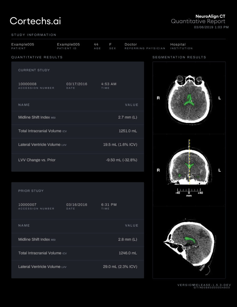
One of the standout features of NeuroAlign CT is its ability to automatically generate quantitative reports that include measures of ventricular volume, intracranial volume, midline shift, and change over time if a prior exam exists.

An axial series with color-coded segmentation of the CSF spaces is also provided, with green denoting CSF in the lateral ventricles. The color-coded images may be used to confirm that the automated segmentation has correctly identified CSF in the ventricles. The lateral ventricular volume (LVV) only includes voxels with CSF density, so volume of blood or a mass in the ventricle (e.g., calcification), will not count towards the total volume measured by NeuroAlign CT.

Thanks to advancements in imaging analytics and AI-driven automation, these volumetric and morphometric measures can now be rapidly quantified from non-contrast CT scans—offering clinicians a real-time window into intracranial pathology. By combining speed and precision, NeuroAlign CT helps radiologists respond quickly and confidently in time-sensitive situations, ultimately enhancing diagnostic confidence, efficiency, and accuracy.
References
[1] Hacking C, Campos A, Alhusseiny K, et al. Midline shift. Reference article, Radiopaedia.org https://doi.org/10.53347/rID-37110


