Structured reporting is transforming radiology by improving consistency, clarity, and interoperability. DICOM SR (Structured Reporting) allows imaging data and associated measurements to be captured in a standardized, machine-readable format—eliminating the need for manual entry and reducing errors. With NeuroQuant® now supporting DICOM SR, radiology teams can achieve tighter integration with PACS, structured reporting systems, and downstream analytics tools.
What is DICOM SR?
DICOM SR is a standardized method for encoding and sharing imaging measurements and diagnostic content alongside the original images. Unlike traditional free-text reports, DICOM SR stores structured clinical observations—volumes, regions of interest, laterality, timestamps—in a format that software systems can interpret and exchange.
This improves data utility in multi-disciplinary environments, enhances longitudinal tracking, and supports research, billing, and AI-driven insights.

DICOM SR vs HL7: Understanding the Difference
While both DICOM SR and HL7 are standards for health data exchange, they serve different purposes:
- DICOM SR focuses on imaging data and structured content generated from medical images. It is ideal for image-derived measurements and annotations.
- HL7 is broader and used primarily for administrative, scheduling, and clinical documentation systems like EHRs.
In practical terms, DICOM SR is optimized for radiology, while HL7 is optimized for hospital-wide communication.
NeuroQuant + DICOM SR: What It Means for You
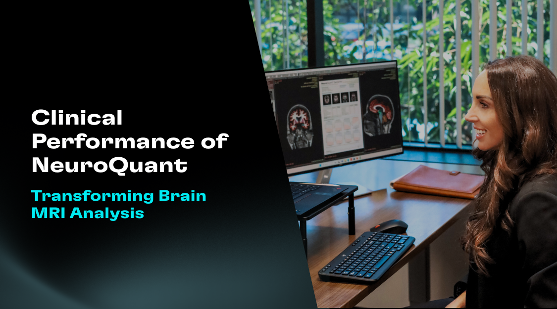
NeuroQuant® now outputs key volumetric data in DICOM SR format, providing precise measurements of brain regions segmented on MRI scans. These values are automatically structured and encoded within the DICOM ecosystem, making them easily accessible and interoperable across systems—independent of any PDF or third-party report format the site may be using. That means:
- No more copying/pasting values from PDF reports
- Reduced risk of transcription errors
- Ready compatibility with structured reporting tools
- Simplified integration into PACS and EHR workflows
This upgrade supports more efficient reporting, stronger communication with referring physicians, and improved data standardization across imaging networks.
Integrating with Other Systems
NeuroQuant’s DICOM SR output is designed for interoperability. Institutions can route DICOM SR data to:
- Structured reporting systems (e.g., PowerScribe, Syngo.via)
- PACS and viewers that support DICOM SR overlays
- Research databases or AI platforms for secondary analysis
Integration is straightforward—your existing DICOM routing rules can often be reused, and the structured report can be queried or displayed alongside standard DICOM images.
Conclusion
With the adoption of DICOM SR, NeuroQuant® takes another step toward streamlining radiology workflows and enhancing clinical communication. Structured, standardized output empowers healthcare professionals to deliver more consistent reports, reduce errors, and drive efficiency in multidisciplinary care.
As imaging becomes more data-driven, tools like NeuroQuant® with DICOM SR support pave the way for smarter, more connected radiology.


