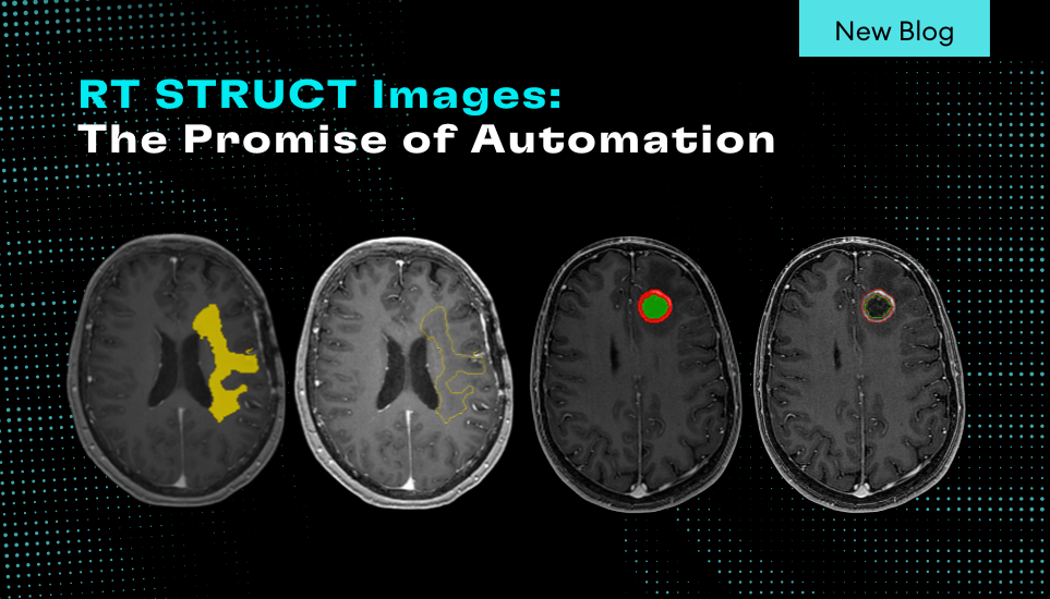
In the realm of neuro-oncology, precision is everything. From diagnosis to treatment planning, clinicians rely on detailed imaging data to make informed decisions that can significantly impact patient outcomes. One of the most critical components in this workflow is the RT STRUCT image, a specialized DICOM format that encapsulates anatomical contours used in radiation therapy planning. Automating the generation of RT STRUCT images with NeuroQuant Brain Tumor can streamline workflows and elevate the standard of care.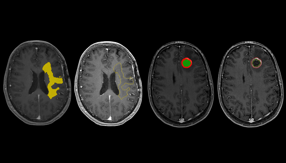
What Are RT STRUCT Images?
RT STRUCT (Radiotherapy Structure Set) images are part of the DICOM-RT standard, designed to support radiation therapy planning. These files contain contours of anatomical structures and regions of interest (ROIs)—such as tumors, organs at risk (OARs), and other critical brain regions—that are essential for:
- Target volume delineation
- Dose calculation and optimization
- Treatment verification and monitoring
In brain tumor cases, RT STRUCT images help radiation oncologists precisely define the tumor boundaries and surrounding healthy tissue, ensuring that radiation is delivered accurately while minimizing damage to critical structures.
How RT STRUCT Images Are Used in Treatment Workflows
The typical workflow for brain tumor treatment using RT STRUCT images involves:
- MRI Acquisition: High-resolution scans are obtained to visualize the tumor and surrounding anatomy.
- Manual Segmentation: Radiologists or dosimetrists manually contour the tumor and OARs—a time-consuming and subjective process.
- RT STRUCT Creation: These contours are saved in RT STRUCT format and imported into treatment planning systems (TPS).
- Radiation Planning: Oncologists use these contours to design radiation plans that maximize tumor coverage and minimize exposure to healthy tissue.
This process, while effective, is labor-intensive and prone to variability between clinicians.
The Challenge: Manual Contouring
Manual segmentation of brain tumors and critical structures is not only time-consuming but also introduces inter- and intra-observer variability. This can lead to inconsistencies in treatment planning and potentially affect patient outcomes. Moreover, the increasing volume of neuro-oncology cases places additional strain on radiology and oncology departments.
The Solution: Automated RT STRUCT Generation with NeuroQuant Brain Tumor
NeuroQuant Brain Tumor offers a powerful solution: automated segmentation of brain tumors and key anatomical structures from MRI scans. By integrating RT STRUCT export capabilities, NeuroQuant can:
- Automatically generate RT STRUCT files with precise contours of tumors and surrounding anatomy.
- Reduce contouring time from hours to minutes, freeing up valuable clinician time.
- Improve consistency and reproducibility across treatment plans.
- Enable faster treatment initiation, which is critical for aggressive or fast-growing tumors.
Clinical Impact and Future Potential
Automating RT STRUCT generation not only enhances operational efficiency but also supports better clinical outcomes. With standardized, high-quality contours, radiation plans can be optimized more effectively, reducing the risk of under-treatment or collateral damage. Additionally, automated RT STRUCTs can facilitate multi-disciplinary collaboration, allowing radiologists, oncologists, and surgeons to work from a common anatomical reference.
As AI-driven tools like NeuroQuant Brain Tumor continue to evolve, the integration of automated RT STRUCT generation represents a significant step forward in personalized, data-driven neuro-oncology care.
