Determining Brain Age Using Machine Learning Combined With Automated Brain Segmentation and PET imaging In Normal, Alzheimer’s Disease and Mild Cognitive Impairment Subjects
Cortechs.ai’s abstract titled “Determining Brain Age Using Machine Learning Combined With Automated Brain Segmentation and PET imaging In Normal, Alzheimer’s Disease and Mild Cognitive Impairment Subjects” was accepted by the Radiological Society of North America.
Lead author and Cortechs.ai Principal Scientist, Weidong Luo, will present his findings at the AI Community Learning Center, Station #1 on December 5, from 12:15 – 12:45 pm.
In addition to two other presentations throughout the conference, you can find the CorTechs team at booth numbers 4055 in South Hall, Level 3 and 10514F in the AI Showcase, Level 2.
Findings
| PURPOSE | |||
| Understanding brain aging may be critical to identify potential biomarkers in neurodegenerative diseases. Here we developed a machine learning based age-prediction model for metabolic and volumetric changes of normal brain structures and evaluated this model on imaging data from patients with Alzheimer’s disease (AD) and mild cognitive impairment (MCI), as well as normal subjects. | |||
| METHOD AND MATERIALS | |||
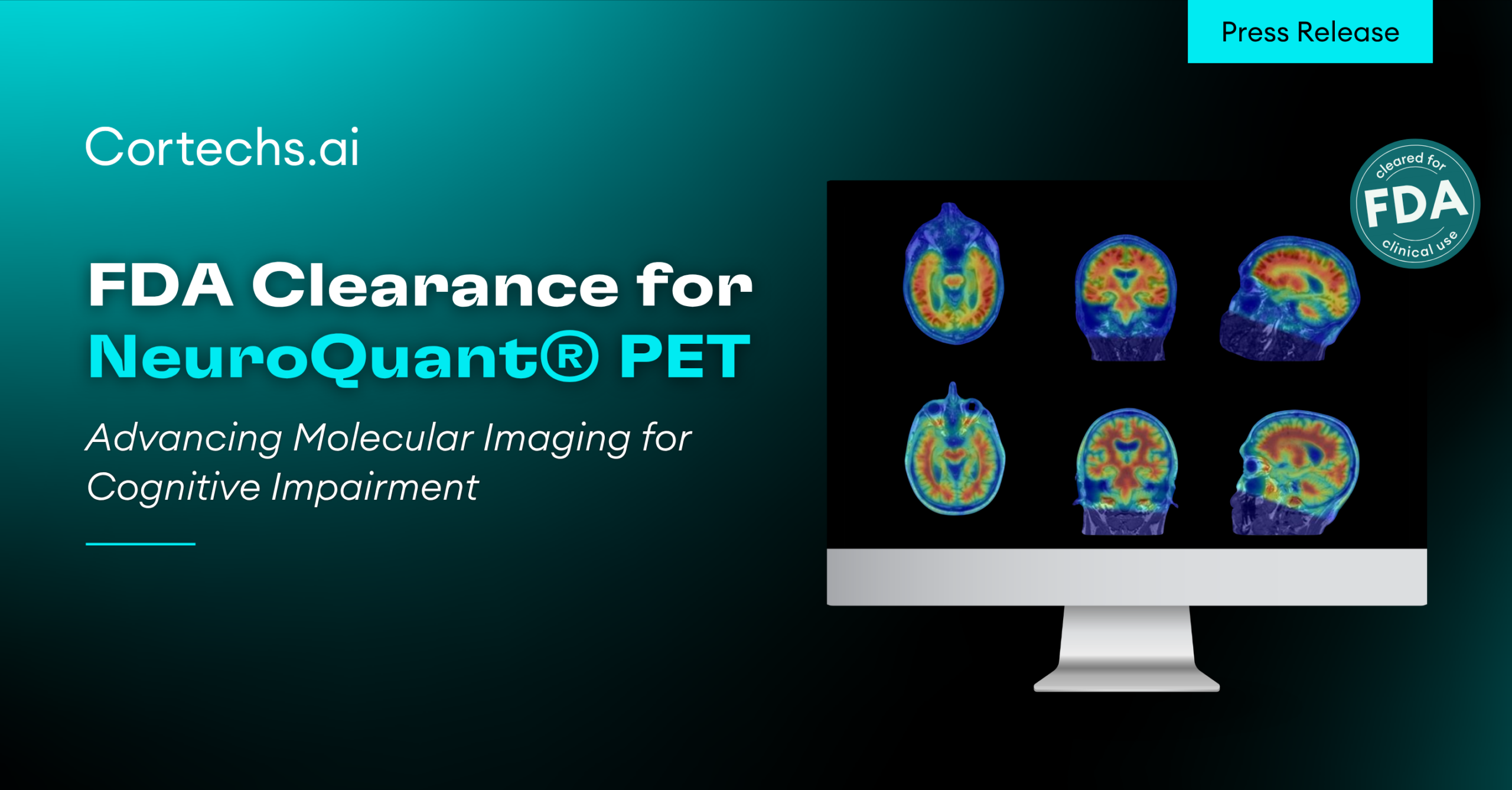
| F18-FDG PET scans and paired T1 weighted MRI brain scans for normal subjects (59-96yo, 157m/169f), MCI (56-93yo, 263m/163f) and AD (56-91yo, 121m/80f) from the Alzheimer’s Disease Neuroimaging Initiative (ADNI) imaging studies were used. All imaging data were processed by an automated brain PET activity analysis and MRI volumetric segmentation tool, PETQuant. The volumetric measurements, as well as the mean SUVR (Standardized uptake value (SUV) ratio) to the brain stem for all brain structures, were calculated. A subset of the normal test data (n=50) was reserved for testing. A brain aging model was created using a Random Forest machine learning algorithm with the normal subjects’ data. MCI and AD data were evaluated using this normal brain aging model. | |||
| RESULTS | |||
| We found the best age prediction was achieved by combining both PET and MRI measurements. For normal subjects, this model showed a high accuracy with the mean difference of -0.23 ± 3.1 years between actual and predicted age by model. In comparison, the MCI group had a mean difference of -2.12 ± 5.13 years, while the AD group had a mean difference of -3.59 ± 5.76 years. The AD subjects younger than 75 years old had the largest disparity between the actual and predicted ages. The predicted age based on PET/MRI measurements was on average 9.35 ± 4.52 year older than the actual age, while AD patients 75 years and above only showed a mean difference of -0.3 ± 3.26 years. | |||
| CONCLUSION | |||
| Our brain aging model showed a significant predicted age difference between normal and younger (<75yo) AD subjects. This suggests that accelerated brain aging could serve as an early biomarker for Alzheimer’s disease. This might help clinicians to identify younger subjects with AD and monitor their disease progression rates. | |||
| CLINICAL RELEVANCE/APPLICATION | |||
| Early diagnosis of Alzheimer’s disease and monitoring disease progression to improve patient management. |
Authors: W Luo, PhD, San Diego, CA; A M Ulug, PhD; W Thompson, PhD; R Haxton; S Magda, PhD; L J Kjonigsen, MSc; et al. (wluo@cortechs.ai)