The team at Cortechs.ai is always looking to further educate ourselves about how brain volumetric imaging is used in the assessment of neurodegenerative conditions. Recently, a practicing Clinical Neuropsychologist visited our San Diego headquarters and gave a very interesting presentation about Temporal Lobe Epilepsy (TLE), its treatments, and how volumetric MRI is involved in the diagnosis.
Seizures are common, and having a single seizure does not mean a person has epilepsy. 4% of the population will have a single seizure at some point in their lifetime. Epilepsy occurs when someone has multiple recurring and unprovoked seizures. 2.2 million people are living with epilepsy in the U.S., and about 150,000 are newly diagnosed per year.
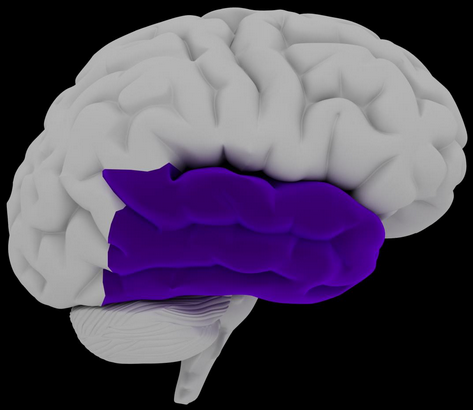
Of the various forms of epilepsy, Temporal Lobe Epilepsy (TLE) is the most common (65% of all cases). For 20-30% of people living with TLE, anti-seizure medications do not work. Physicians and researchers haven’t yet pinpointed what causes TLE. They do know, however, that certain aspects might lead to epilepsy in the future, such as a history of febrile convulsions under the age of 5 and a family history of epilepsy. Early intervention can improve the quality of life for people with TLE. Surgery can be an effective treatment option, especially for those where the source of the seizures is clear and where the medicines don’t control the seizures.
TLE is localized in the temporal lobe of one side of the brain with the hippocampus being the most common structure affected. Diagnosis of TLE is made using an electroencephalogram (EEG), which detects electrical activity in the brain using electrodes attached to the scalp. A MRI scan of the brain is typically performed, to give physicians better visualization on the brain and help determine which structure on which side of the brain may have structural damage, and thus could be the source of the seizures.
NeuroQuant® is a leading medical device software that is used in post-processing of the MRI scan. It maps the sub-cortical brain structures and provides their volumetric imaging. NeuroQuant uses the images from the MRI scan to measure the volume of certain brain structures and the asymmetry between them, in this case the left and right hippocampi, providing important information in the assessment of temporal lobe epilepsy. For TLE, the NeuroQuant Hippocampal Volume Asymmetry Report can be used to help assess which side of the hippocampus may have damage and thus could be causing the seizures, especially in subtle instances.
