The ability to compare the volumes of patients’ brain structures to brain structure volumes of healthy controls is a powerful tool to help physicians assess possible existence of neurodegeneration or the progression of a neurodegenerative disorder.
To adjust for any variation in brain size, sex difference, and inter-individual head-size in a population, brain structure volumes are normalized (or scaled) to a common measurement.
Cortechs.ai’ quantitative imaging software uses Intracranial Volume (ICV) to normalize an individual’s brain structure volumes, and automatically calculates it during the post-processing of brain MRIs for volumetric analysis. This consistent measure gives clinicians additional context of their patient’s brain structure volumes, regardless of head size.
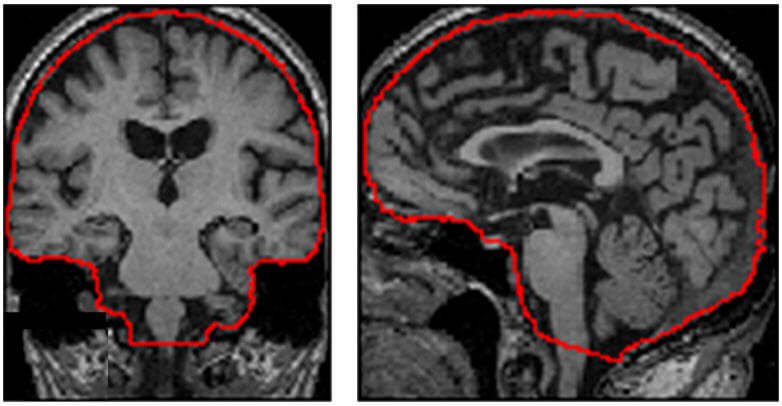
ICV, also known as TIV (Total Intracranial Volume), is the volume of the cranial cavity as taken from a 3D T1 MRI, as outlined by the supratentorial dura matter, or cerebral contour when dura is not clearly detectable.

ICV is a very consistent measure across the lifespan of a patient, making it a reliable tool for correction of head size variation across individuals. This correction is especially helpful when assessing neurodegenerative conditions.
For example, Patient A and Patient B both have thalamic volumes that are consistent with the average for their age and sex-matched population. Patient A is 5’6” tall, with an average head circumference, while Patient B is 6’2” tall with an above average head circumference. If the thalamic volumes could be interpreted without correcting for ICV, both patients would yield a normal result. However, once the patient’s thalamic volumes are normalized to their individual ICV, it could become apparent that Patient B’s thalamic volume is lower than expected for their age and gender.
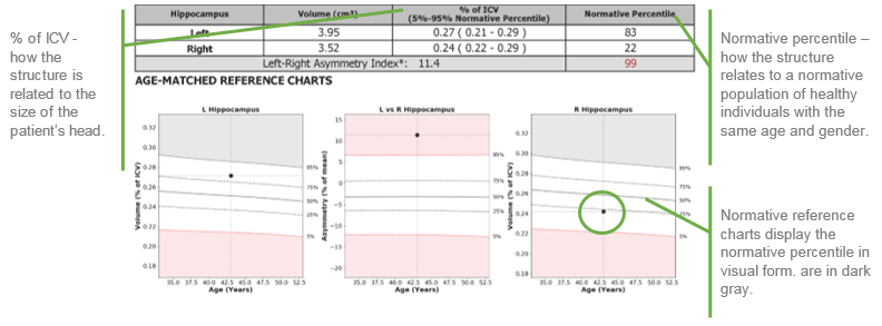
NeuroQuant reports provide brain structure volumes (relevant to the report), % ICV and normative percentiles. NeuroQuant automatically calculates the ICV to accurately compare an individual’s brain structure volumes to Cortechs.ai’ age and sex-matched normative reference data. The ICV normalization and reference comparison on NeuroQuant reports provide physicians with a framework to better understand brain structure volumes when assessing patients.
Need more information about ICV or Cortechs.ai quantitative image products? Contact us.


