Quantitative MR volumetry in temporal lobe epilepsy

Neuroradiology: quantitative MR volumetry in temporal lobe epilepsy

Quantitative MR imaging using NeuroQuant® can enhance standard visual analysis, providing a viable means for translating volumetric analysis into clinical practice and increasing the detection of hippocampal atrophy in temporal lobe epilepsy in both community and tertiary care settings. Radiology August 2012 264:542-550; Published online June 21, 2012.

[button]Download[/button]

More Resources

09/01/2026

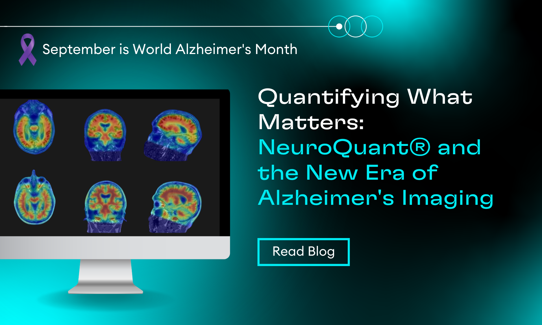
Quantifying What Matters: Advancing Alzheimer’s Imaging with NeuroQuant®

Explore how quantitative imaging is transforming Alzheimer’s care, from structural volumetrics and amyloid PET to treatment monitoring with NeuroQuant®.

08/19/2026

Brain Metastases Imaging Insights from SNO/ASCO 2026

Explore key insights from SNO/ASCO 2026 on brain metastases imaging, including lesion tracking, volumetric analysis, SRS workflows, and automated segmentation.

08/18/2026

Cortechs.ai Announces FDA Clearance for NeuroQuant® PET: Advancing Molecular Imaging for Cognitive Impairment

NeuroQuant® PET receives FDA 510(k) clearance, advancing quantitative PET imaging for cognitive impairment.

08/04/2026

Cortechs.ai Expands European CE Marking Across AI Imaging Platform, Accelerating Global Growth

Cortechs.ai expands EU CE Marking for the latest NeuroQuant®, NeuroQuant® Brain Tumor, and OnQ™ Prostate, advancing global AI imaging innovation

07/29/2026

Read Your Segmentations in the Same Space You Read the Study: Native Space Outputs in NeuroQuant v5.3.0

Review lesion segmentations directly on your original MRI images with NeuroQuant® v5.3.0.

07/23/2026

What AAIC 2026 Told Us About the Future of Dementia Imaging — And Where NeuroQuant Fits

Quantitative MRI brain volumetry with NeuroQuant.
Scroll to Top