Less than perfect positioning
Typically, in every MRI scan, the patient’s head and therefore the brain, is aligned slightly differently. This is primarily dependent on patient positioning, including variability of head rotation and tilt. Manual segmentation of MR images is highly reliant on accurate positioning of the patient’s head and deviations from cardinal alignment increases the observer-based variability, thereby reducing segmentation accuracy.
This is especially concerning when evaluating changes in a follow-up scan, making comparisons to baseline less precise even in side-by-side comparisons.
In addition, MR images are known for not being geometrically accurate due to a large variety of factors impacting the homogeneity of the magnetic field. When adding image distortions on top of the alignment variability, manual segmentation becomes highly subjective and reproducibility requires highly experienced and trained individuals.
Automated image consistency and reproducibility
When using Cortechs.ai’ NeuroQuant® and PETQuant™ products, head alignment and slice presentation are automatically corrected for magnetic field inhomogeneity and non-cardinal patient positioning, providing consistency and reproducibility from scan-to-scan, site-to-site and in follow-up patient scans, without operator intervention. (Example below.)

Correction of spatial distortion
MR images inherently contain spatial distortions due to magnetic field properties of the magnet, magnetic field inhomogeneity introduced by the patient’s body, and inhomogeneous tissue magnetization. Cortechs.ai automatically corrects for these spatial distortions, allowing for improved spatial integrity of the derived images generated by NeuroQuant and PETQuant. (Example below.)
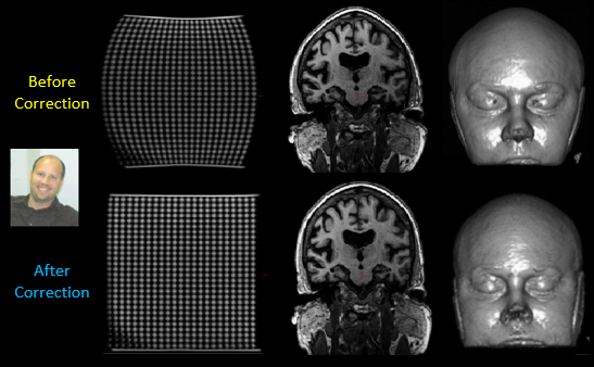
Image quality standardization
When using Cortechs.ai’ products, radiologists receive high image quality standardization through our consistent head alignment and slice presentation as well as our automatic spatial distortion correction. Additional benefits of using Cortechs.ai’ products include:
- Visual identification of sub-cortical brain structures
- Quantification of sub-cortical brain structures
- Comparisons of structures/regions of interest at different time points
- Overlay of images from different modalities


