NeuroQuant®: Contrast agents not required

Factors which degrade the technical quality of MR images or pathologic processes, which significantly affect neuroanatomic structure, may decrease the accuracy of NeuroQuant® output. One of these factors is the use of contrast agents.

Contrast agents are typically used to study vasculature, something that is not needed for volumetric MR imaging. NeuroQuant requires a non-contrasted, 3D T1 sagittal sequence to segment and measure brain structure volumes.

NeuroQuant does not support a contrast enhanced study. The presence of contrast will cause misregistration of cortical and subcortical structures. The segmentation, volumetrics and normative comparisons will be inaccurate when contrast agents are used.

Some examples of NeuroQuant processed with and without contrast agents are shown below.
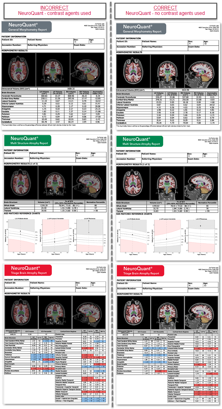
Cortechs.ai | NeuroQuant®: Contrast agents not required

More information about using NeuroQuant can be found here.

More Resources

10/10/2026

World Mental Health Day: Why Brain Health and Mental Health Belong Together

Behind every brain scan is a person. Here’s why their voice matters.

09/30/2026

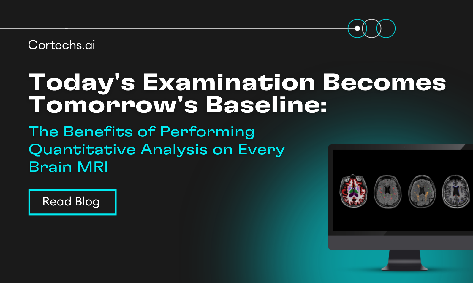
Today’s Examination Becomes Tomorrow’s Baseline: The Benefits of Performing Quantitative Analysis on Every Brain MRI

A single brain MRI shows where a patient is today. A quantitative baseline can help show how the brain changes over time.

09/18/2026

What a Head CT Can and Can’t Tell You About a Concussion

For Concussion Awareness Day, learn how quantitative CT analysis can help measure subtle structural changes and support follow-up after traumatic brain injury.

09/09/2026

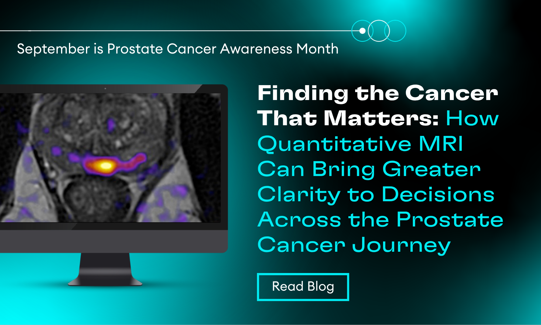
Prostate Cancer Awareness Month: Finding the Cancer That Matters

How quantitative MRI can bring greater clarity to decisions across the prostate cancer journey.

09/01/2026

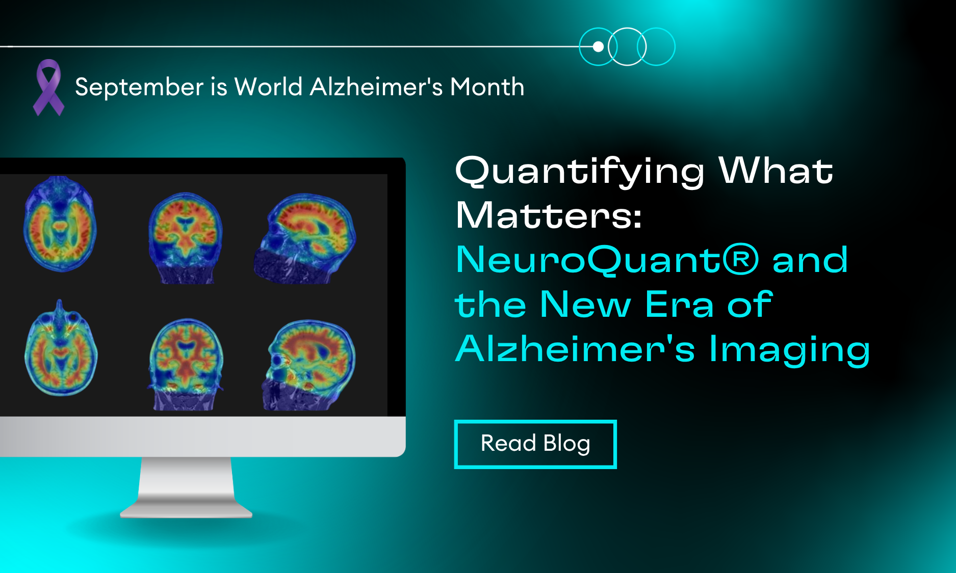
Quantifying What Matters: Advancing Alzheimer’s Imaging with NeuroQuant®

Explore how quantitative imaging is transforming Alzheimer’s care, from structural volumetrics and amyloid PET to treatment monitoring with NeuroQuant®.

08/19/2026

Brain Metastases Imaging Insights from SNO/ASCO 2026

Explore key insights from SNO/ASCO 2026 on brain metastases imaging, including lesion tracking, volumetric analysis, SRS workflows, and automated segmentation.
Scroll to Top