Our COVID-19 Projects Include:

[vc_section]
[vc_empty_space height=”20px”]

Brain Volumetrics

NeuroQuant and LesionQuant | Available for Clinical Use
Introduction

Cortechs.ai is providing the most widely used clinical brain morphometry tool to help with the COVID-19 pandemic. LesionQuant FLAIR Lesion Report, NeuroQuant Triage Brain Atrophy (TBA) and Custom COVID-19 reports will be available, free of charge, to all facilities for COVID-19 patients or for COVID-19 research. As the research on the neurological effects of the disease is ongoing, our offering may be updated accordingly.

Watch the Recorded Webinar: Brain Volumetrics for COVID-19

Complimentary COVID-19 Package

LesionQuant

FLAIR Lesion Report 

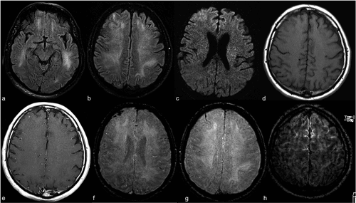
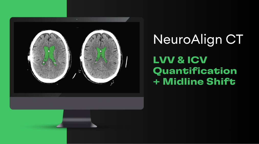
According to recent clinical findings, COVID-19 patients under the age of 50 are often presenting with large vessel strokes. The LesionQuant report has the unique feature of giving detailed volumetric structural brain measurements in addition to lesion volumes, percent of intracranial volume, and total lesion burden. With speed and accuracy, LesionQuant performs automated FLAIR lesion segmentation and quantification along with the following brain regions quantification: whole brain, thalamus, cortical gray matter, and cerebral white matter. It may also be useful in evaluating a brain with microstrokes and for short term care because it can guide more aggressive anticoagulant or antiplatelet therapy. It may also be useful for long term longitudinal tracking to measure drug effectiveness and to determine whether changes in disease-modifying therapies need to be altered.

[vc_images_carousel images=”9232″ img_size=”full” onclick=”link_no” speed=”4000″ autoplay=”yes” wrap=”yes”]

NeuroQuant

TBA Report

The TBA report is included so clinicians can quickly identify swelling in the whole brain, and brain regions, particularly the frontal and temporal lobes as seen in some severe COVID-19 cases. For encephalitis, clinicians can quickly review volumes for the whole brain, individual lobes, and numerous structures relevant to COVID-19 providing an immediate overview from both a global and individual structure perspective.

Custom Report 

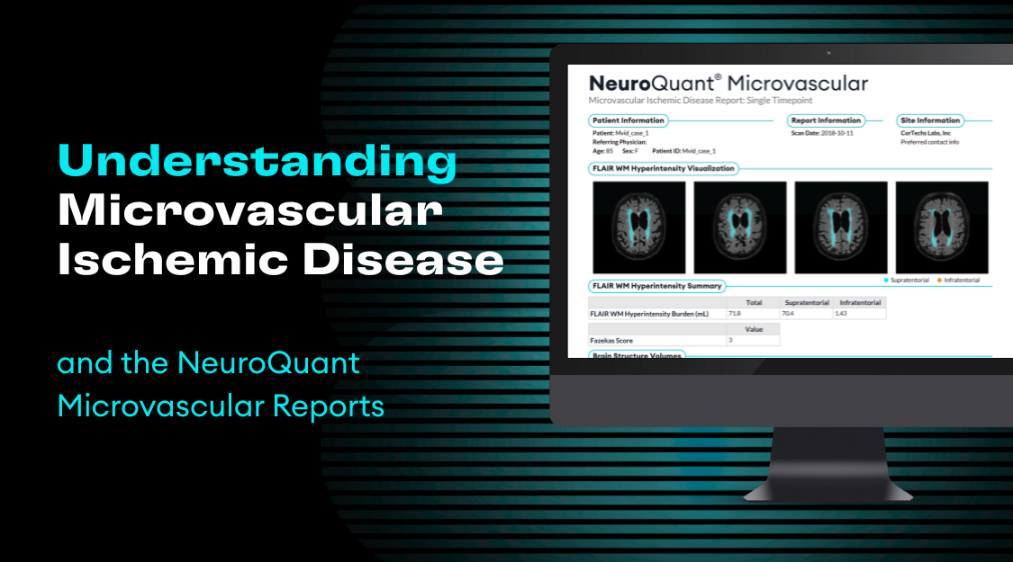
The Custom COVID-19 Report includes brain structures that are potentially affected by COVID-19: cerebellum, brainstem, whole brain, frontal lobes, frontal poles, medial orbitofrontal lobes, anterior middle frontal lobes, thalamus, amygdalae and cerebral white matter hypointensities. This report will show the raw volume, percent of ICV, and normative percentiles for each structure. NeuroQuant’s custom volumetric reports were designed to provide users with flexibility in report design. The predefined COVID-19 custom report is an example of how users can create their own NeuroQuant report with structures that meet their specific needs.

[vc_images_carousel images=”9195,9196,9228″ img_size=”full” onclick=”link_no” speed=”4000″ autoplay=”yes” wrap=”yes”]

The NeuroQuant and LesionQuant reports offered as part of the complementary COVID-19 package are to be used for followup of COVID-19 patients. They are not intended for COVID-19 diagnosis.

LesionQuant’s FLAIR Lesion Report and NeuroQuant’s TBA and Custom Report are free for clinicians and researchers.

Apply for complimentary access to the COVID-19 report package

[/vc_section][vc_section]
[vc_empty_space height=”25px”]

Interpretable AI for COVID-19

Chest X-Ray | In Development 
Clinical Need

Role of Chest Radiographs in COVID-19
American College of Radiology identified the importance of chest x-ray by stating “As COVID-19 spreads in the U.S., there is growing interest in the role and appropriateness of chest radiographs (CXR) for the screening, diagnosis, and management of patients with suspected or known COVID-19 infection.” Compared to CT, CXR requires less personal protective equipment (PPE) and less exposure to hospital staff and other patients. Chest x-ray is fast and available in most acute care settings. More severe radiographic findings are suspected to be associated with a higher risk of intubation and possible death. Accurate interpretation can be challenging, especially in the differentiation of COVID-19 from non-COVID pneumonia.

Timely Care
Early risk stratification and rapid diagnosis are critical to improved COVID-19 patient outcomes. AI can provide insights that can be used to triage the subset of patients most likely to have poorer outcomes that will need a higher level of care, assist with timely care escalation and resource planning.

Second Detection Opportunity in Symptomatic Patients
While PCR and laboratory tests will likely remain the first-line test for detection. Chest x-ray could become a valuable safety net for symptomatic false negatives. In a cohort of 58 individuals, 9% showed chest x-ray abnormalities before eventually testing positive for COVID-19 using lab tests (Wong et al. Frequency and Distribution of Chest Radiographic Findings in COVID-19 Positive Patients. Radiology. 2020).

Insights into Radiographic Signatures of COVID-19
Additional research is needed to precisely define findings on chest imaging in COVID-19 that are specific, and do not overlap with other infections, including influenza and H1N1 and bacterial types of pneumonia. Our region attribution-based saliency approach applied to neural networks can be used to explain the radiographic features that most influence the discrimination of COVID-19. In addition, our approach can be used to better understand the patterns of findings that are associated with clinical deterioration, intubation, and mortality.

[vc_message message_box_color=”grey” icon_fontawesome=”fas fa-angle-double-right”]Available soon for research use.[/vc_message][vc_progress_bar values=”%5B%7B%22label%22%3A%22Development%22%2C%22value%22%3A%2265%22%7D%5D” bgcolor=”bar_blue” units=”%”]
Objective

Create a quick and highly available tool for rapid risk stratification and triage of COVID-19 patients by improving the interpretation of chest radiographs.

Methodology

Cortechs.ai, in conjunction with multiple academic partners including URMC, has developed an AI-based algorithm using deep learning to provide rapid diagnostic decision support for COVID-19 on chest x-rays. Our algorithm has achieved a ROC-AUC of >0.95 on a preliminary validation set in the discrimination of COVID-19 versus non-COVID-19 related pneumonia on CXR. We then utilize a probability-weighted region-based attribution saliency method to identify signatures that have the most influence on the neural network’s prediction, yielding insights into the contributing radiographic features that are indicative of COVID-19 pneumonia. In a case-based preliminary evaluation, highlighted regions and radiograph features corroborated radiographic findings such as patchy consolidations, ground-glass opacities, and perihilar infiltrates that are frequently associated with COVID-19 infection as well as more severe COVID-19 outcomes such as mortality and intubation.

The potential for this technology to become clinically significant increases when the interpretable AI is evaluated for its utility in the prediction of clinical deterioration, intubation, and mortality.

COVID-19 Pneumonia

Probability of COVID-19 = 0.90
Probability of other pneumonia = 0.01

COVID-19 Saliency Overlay
Raw X-Ray

Region-based attribution map highlights the radiographic features most indicative of COVID-19 for the neural network’s prediction for this confirmed COVID positive patient. Notable features highlighted include patchy consolidations, perihilar distribution, and peripheral distribution.

[vc_btn title=”Predict COVID-19 from X-Ray” color=”primary” size=”lg” align=”center” i_icon_fontawesome=”fas fa-external-link-alt” button_block=”true” add_icon=”true” link=”url:https%3A%2F%2Fcortechs.ai%2Fcovid-19-interpretable-ai-demo%2F||target:%20_blank|”]
[vc_message]Participate in this project
  • Clinicians working in acute care settings (e.g. ED, ICU) and have clinical risk factors (e.g. EMR data, labs) and chest x-ray data
  • Clinical collaborator working in an acute care setting interested in evaluating our application
  • Researchers interested in investigating radiographic signatures of COVID-19 on chest x-ray and their correlation with clinical outcomes
Click here to participate [/vc_message]
[/vc_section][vc_section]
[vc_empty_space height=”25px”]

Intelligent Integration of Genetic and Clinical Risk Factors

In Development 
COVID-19 Integrated Risk Report

The COVID-19 IntegratedRisk Report predicts a patient’s risk for severe COVID-19 infection using polygenic risk scores for eight blood markers and basic health information such as (age, sex, comorbidities). Previously Gong et al. have shown that these blood traits are predictors with severe COVID-19 outcomes (e.g. Acute Respiratory Distress Syndrome ARDS). Generally, it is known that these genetics-based estimates of blood traits are robustly associated with susceptibility to respiratory infections (Tanigawa and Rivas 2020).

Future work including investigating the utility of our approach in more acute settings with the addition of clinical variables includes a patient’s symptoms, vitals, demographics, comorbidities, routine lab tests, and AI-assisted chest X-ray interpretation.

  • Risk Assessment

    Personalized identification of individuals at high risk for developing severe COVID-19 illness.

  • Better Care/Treatment

    Identifying high-risk patients will enable earlier treatments and better allocation of limited hospital resources.

  • Identify individuals who have a risk to experience severe symptoms if contracted

    Understand the risk of severe outcomes if a patient were to become infected.

[vc_message message_box_color=”grey” icon_fontawesome=”fas fa-angle-double-right”]Available soon for research use.[/vc_message][vc_progress_bar values=”%5B%7B%22label%22%3A%22Development%22%2C%22value%22%3A%2235%22%7D%5D” bgcolor=”bar_blue” units=”%”]
[vc_message]Participate in this project

If you are a clinician and treating COVID-19 patients in an acute care setting, click here to participate. [/vc_message]

[/vc_section][vc_section disable_element=”yes”]
[vc_empty_space height=”25px”]

Genetic Risk

COVID-19 Genetic Risk Report

The COVID-19 GeneticRisk Report uses a patient’s genetics to report their ABO blood type and quantitative measurements of 5 different blood markers that are clinically relevant to the severity of COVID-19 infections. The blood markers include monocytes, eosinophils, lymphocytes, neutrophils, and white blood cells. This report is based on published polygenic risk scores from Tanigawa and Rivas (2020).

The turnaround time for obtaining genetics is currently too long (4-5 days through Diagnomics) for the acute care setting.

[vc_message message_box_color=”grey” icon_fontawesome=”fas fa-exclamation-circle”]Internal research only. No direct distribution.
Contact us for partnership opportunities.[/vc_message][vc_progress_bar values=”%5B%7B%22label%22%3A%22Development%22%2C%22value%22%3A%2225%22%7D%5D” bgcolor=”bar_blue” units=”%”]
  • Identify individuals who have a risk to experience sever symptoms if contracted

    Understand the risk for severe outcomes if a patient were to become infected.

[vc_images_carousel images=”9082″ img_size=”full” onclick=”link_no” speed=”4000″ autoplay=”yes” wrap=”yes”]
[vc_message]Participate in this project

We seek data to validate associations between genetic-based blood measurements and COVID-19 severity. If you have tested positive for COVID-19 and have participated in 23andMe or Ancestry.com genetic testing, please fill out this form[/vc_message]

[/vc_section]