Improving early and accurate diagnosis of Alzheimer’s disease, the most common form of dementia
Early and accurate diagnosis of Alzheimer’s disease can benefit both patients and caregivers by allowing them more time to develop a plan and explore different interventions such as clinical trials, which may slow disease progression. Alzheimer’s disease (AD) is the most common neurodegenerative disease and form of dementia globally. It develops when neuronal atrophy occurs, in both white and grey matter[2]. Neuronal or cerebral atrophy is the loss or weakening of neuronal connections as a result of neurons in brain tissue becoming injured or dying[3]. As atrophy occurs, brain regions, especially hippocampi shrink, lateral ventricles expand, and there is a significant loss of brain volume[3]. Atrophy impacts many different aspects of life and brain function, causing difficulty communicating, inability to recollect memories and information, and impaired physical control[1].
More than six million Americans are currently living with Alzheimer’s disease[4]. As research for a cure continues, the death rate of those living with Alzheimer’s has increased by 145 percent, killing more than breast cancer and prostate cancer combined.
Identifying AD in its earliest stage, mild cognitive impairment (MCI), allows the physician to monitor progression and provides the patient the ability to try different lifestyle changes that may prolong progression.
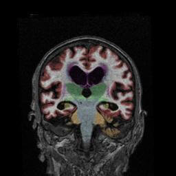
Physicians use several evaluation tools to make an accurate assessment of memory loss in patients, one of them being a magnetic resonance imaging (MRI) scan of the patient’s brain. A NeuroQuant analysis can be part of a routine physician-prescribed MRI procedure.
NeuroQuant, the first FDA 510(k) cleared medical software for quantitative analysis of brain MRI, provides brain structure volumetrics to assist physicians in fast and accurate diagnoses of neurodegenerative diseases.
When assessing dementia, NeuroQuant uses images from an MRI scan to measure the volume of specific brain structures imperative to the evaluation of memory loss. The software allows physicians to pinpoint specific areas of concern, removing the subjectivity of an MRI scan.
The NeuroQuant Dementia report is most applicable when assessing a case of memory loss.
Volumetrics provided on the NeuroQuant Dementia report include a hippocampal occupancy score, volumes, percent of intracranial volume, and normative reference percentiles for the hippocampi, entorhinal cortex, superior lateral ventricles, and inferior lateral ventricles.
The following case study reviews a patient with reported memory loss who underwent a NeuroQuant volumetric MRI analysis.
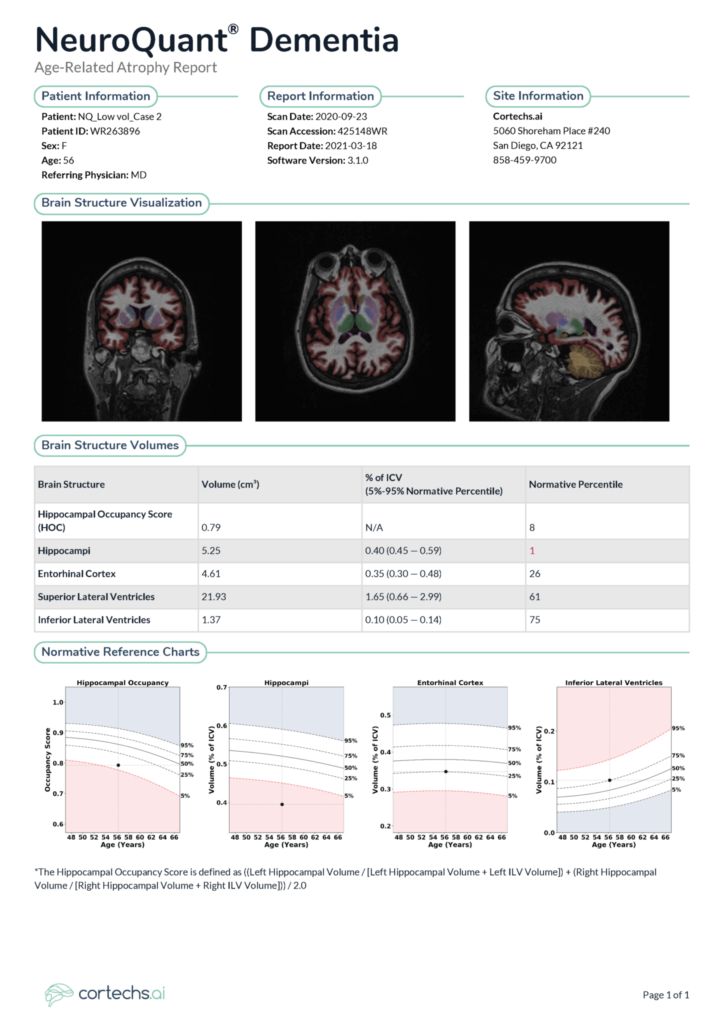
NeuroQuant Findings
The hippocampal occupancy score measures 0.52 cm3, which is over 2 standard deviations below the mean (1%) and therefore statistically significant for the patient’s age and ICV. The volume of the hippocampi measures 5.55 cm3, which is over 2 standard deviations below the mean (1%) and therefore statistically significant for the patient’s age and ICV.
The volume of the entorhinal cortex measures 4.12 cm3 (2%) which is statistically significant for patient age.
The volume of the superior lateral ventricles measures 63.88 cm3 which is more than 2 standard deviations above the mean (99%) and therefore statistically significant for patient age. The volume of the inferior lateral ventricles measures 5.40 cm3 which is more than 2 standard deviations above the mean (99%) and therefore statistically significant for patient age.
Sample radiologist’s impression
Analysis demonstrates statistically significant hippocampal atrophy and statistically significant compensatory lateral ventricle enlargement. There is also a statistically significant reduction of the entorhinal cortex and the HOC, all of which are highly associated with progression from mild cognitive impairment to Alzheimer’s disease.
NeuroQuant 3.1
NeuroQuant version 3.1 was released without modifications to the products’ volumetric algorithm, and volumetric measurements of this release are identical to those computed in the 3.0.2 and 3.0.0 releases.
NeuroQuant 3.1 is now available to current customers, both cloud-based and installed. There is no automatic migration to NeuroQuant 3.1 and users must coordinate with their account manager to activate this version.
To learn how you can add NeuroQuant to your dementia imaging workflow, schedule a discovery call.
References
- Alzheimer’s Disease and the Neuroscience of Aging | National Institute on Aging. (n.d.). National Institute on Aging; https://www.nia.nih.gov/about/budget/alzheimers-disease-and-neuroscience-aging-3
- Toepper M. (2017). Dissociating Normal Aging from Alzheimer’s Disease: A View from Cognitive Neuroscience. Journal of Alzheimer’s disease : JAD, 57(2), 331–352. https://doi.org/10.3233/JAD-161099
- What Happens to the Brain in Alzheimer’s Disease? | National Institute on Aging. (n.d.). National Institute on Aging;. https://www.nia.nih.gov/health/what-happens-brain-alzheimers-disease
- (2021). 2021 Alzheimer’s Disease Facts And Figures [pdf]. Alzheimer’s association. https://www.alz.org/media/Documents/alzheimers-facts-and-figures-infographic.pdf
