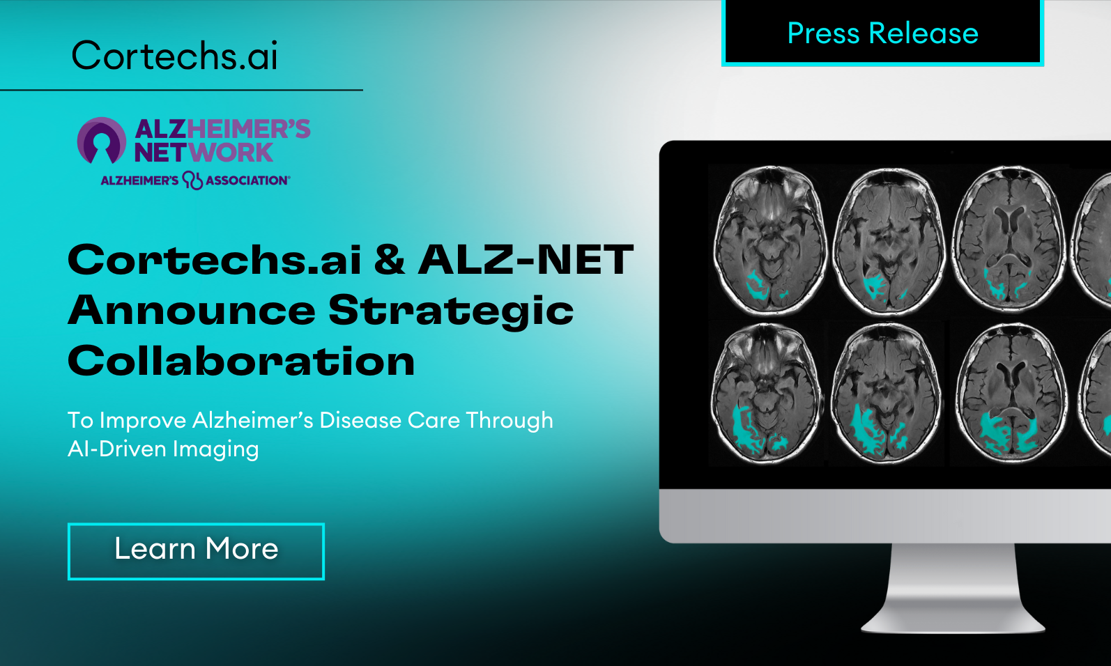
Traumatic Brain Injury (TBI) is a serious public health concern, affecting millions of individuals annually, including athletes, military personnel, and accident victims. While some TBIs, such as concussions, may resolve over time, others can lead to persistent neurological consequences. Severe TBIs, in particular, can result in chronic brain atrophy, network dysfunction, and long-term cognitive, emotional, and motor impairments. Without proper monitoring, these injuries may contribute to progressive neurodegeneration, impacting quality of life and increasing the risk of conditions such as chronic traumatic encephalopathy (CTE).
Traditional TBI assessment relies heavily on clinical evaluations and symptom checklists, which are valuable for immediate TBI assessment but can be subjective and unable to detect underlying structural damage. Advanced MRI neuroimaging with NeuroQuant, an AI-driven volumetric analysis software, provides a more precise and objective approach to evaluating brain injuries. This technology helps clinicians and researchers visualize structural damage, quantify brain volume changes, and track recovery over time—critical components in guiding treatment and rehabilitation decisions.
Tracking TBI Recovery with NeuroQuant
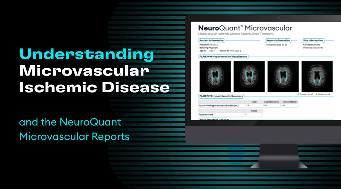
One of the most powerful tools for longitudinal TBI monitoring is NeuroQuant, an FDA-cleared automated software that analyzes MRI scans to quantify brain volume. The Total Brain Atrophy (TBA) Report within NeuroQuant provides key metrics that can guide TBI assessment, including:
- Quantitative volumetric data for brain regions commonly affected by TBI (e.g., hippocampus, thalamus, brainstem)
- Color-coded analysis that highlights areas of atrophy and structural changes
- Normative database comparison to track deviations from healthy brain volumes
- Longitudinal tracking to monitor recovery progress and detect emerging neurodegeneration
By using NeuroQuant’s objective imaging data, clinicians can track changes over time and personalize rehabilitation plans.

The Role of Advanced Imaging in TBI Recovery
Research in the field of neuroimaging has demonstrated the long-term impact of severe TBIs on brain structure. Studies tracking TBI patients over extended periods have shown significant brain volume changes post-injury, particularly in regions associated with cognitive function. These findings emphasize the importance of advanced MRI techniques in identifying chronic-stage pathological changes, such as encephalomalacia and focal or diffuse atrophy, which can persist well beyond the acute phase of injury.
As MRI and AI-driven analyses continue to evolve, they enable researchers and clinicians to detect subtle, long-term alterations in brain volume and connectivity. When integrated with neuropsychological assessments, these tools help correlate structural changes with functional impairments, providing a comprehensive view of TBI recovery. This integration is particularly valuable for assessing the extent of neurological complications and predicting long-term recovery trajectories.
Conclusion: A Data-Driven Approach to TBI Recovery
The evolution of AI-driven MRI analysis is revolutionizing TBI assessment and recovery monitoring. By leveraging tools like NeuroQuant, clinicians can move beyond subjective symptom evaluations and implement data-driven, personalized treatment strategies.TBI recovery is a highly individualized process, and no two patients will experience the same trajectory. Long-term monitoring with volumetric analysis is essential to detect delayed atrophy or secondary neurodegeneration, evaluate brain plasticity and potential for functional recovery. For athletes, military personnel, and anyone recovering from a TBI, these innovations offer hope for better outcomes, safer return-to-play decisions, and improved long-term brain health.