Quantitative MR volumetry in temporal lobe epilepsy

Neuroradiology: quantitative MR volumetry in temporal lobe epilepsy

Quantitative MR imaging using NeuroQuant® can enhance standard visual analysis, providing a viable means for translating volumetric analysis into clinical practice and increasing the detection of hippocampal atrophy in temporal lobe epilepsy in both community and tertiary care settings. Radiology August 2012 264:542-550; Published online June 21, 2012.

[button]Download[/button]
More Resources

11/10/2026

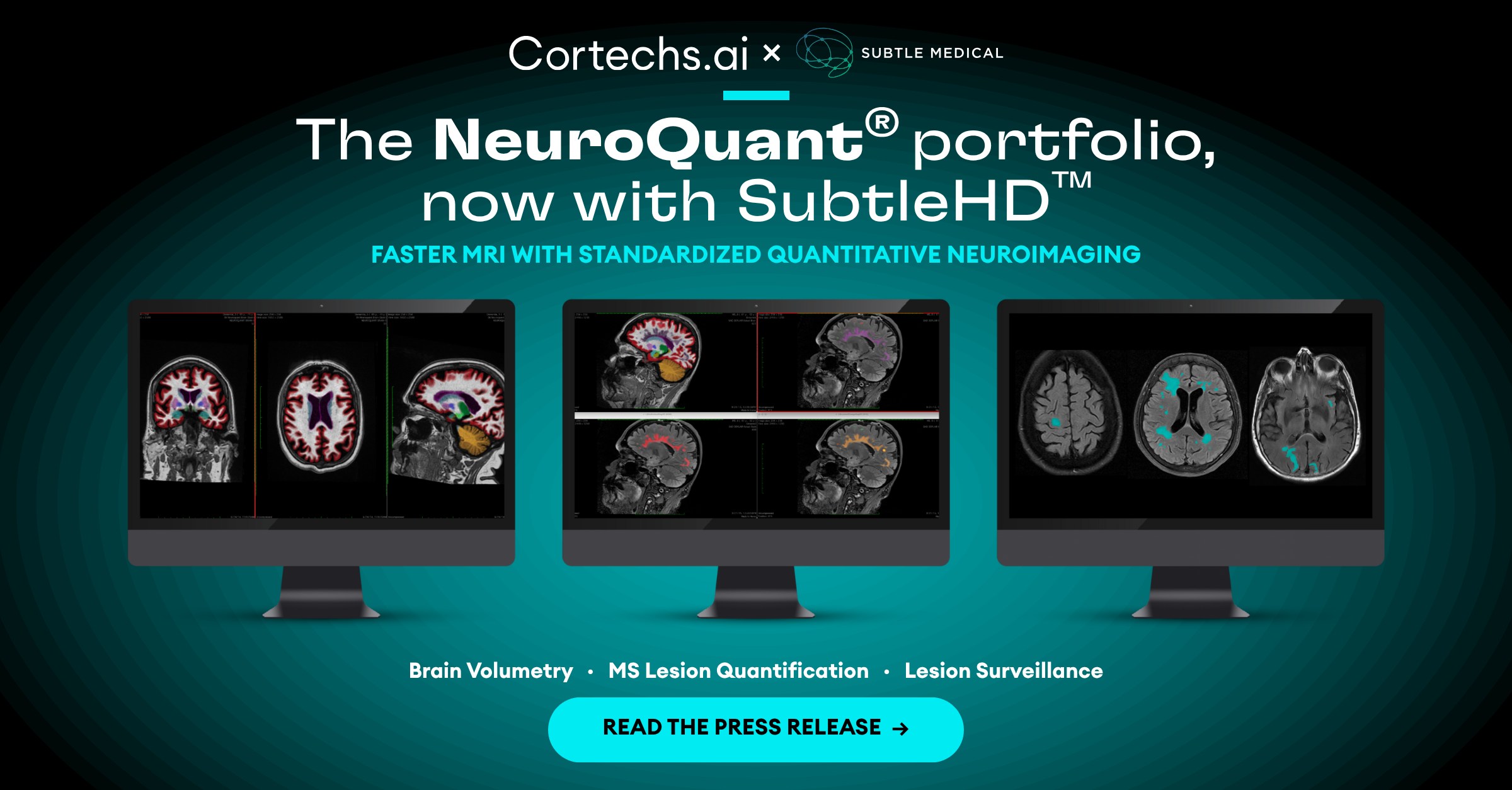
Cortechs.ai and Subtle Medical Partner to Deliver Faster MRI with Standardized Quantitative Neuroimaging

Cortechs.ai and Subtle Medical partner to accelerate MRI scans and deliver standardized, AI-powered quantitative neuroimaging.

10/30/2026

What to Expect at the Cortechs.ai Booth at RSNA 2026

Visit Cortechs.ai at RSNA 2026, Booth 5107. See Cortechs Connect, FDA-cleared NeuroQuant PET and live demos of our AI quantitative imaging tools.

10/14/2026

Cortechs.ai and CARPL.ai Partner to Expand Global Access to AI-Powered Imaging Solutions

Cortechs.ai joins CARPL.ai’s enterprise AI ecosystem to expand access to quantitative imaging solutions worldwide.

10/10/2026

World Mental Health Day: Why Brain Health and Mental Health Belong Together

Behind every brain scan is a person. Here’s why their voice matters.

09/30/2026

Today’s Examination Becomes Tomorrow’s Baseline: The Benefits of Performing Quantitative Analysis on Every Brain MRI

A single brain MRI shows where a patient is today. A quantitative baseline can help show how the brain changes over time.

09/18/2026

What a Head CT Can and Can’t Tell You About a Concussion

For Concussion Awareness Day, learn how quantitative CT analysis can help measure subtle structural changes and support follow-up after traumatic brain injury.
Scroll to Top