
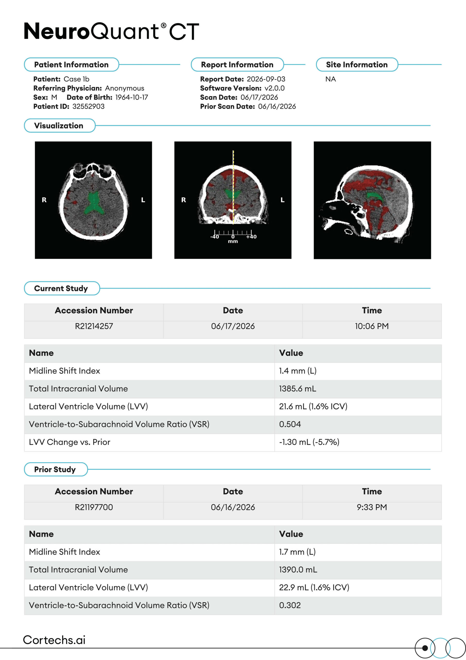
Patient, Report, Referring Physician, and Site Information, including software version and current and prior scan dates.
Visualization: Color-coded CT images in the axial, coronal, and sagittal planes highlighting the lateral ventricles and subarachnoid CSF spaces, with a midline reference and millimeter scale on the coronal view.
Current Study: Accession number, date, and time of the current CT exam, with measurements for midline shift index, total intracranial volume, lateral ventricle volume, ventricle-to-subarachnoid volume ratio, and change in lateral ventricle volume vs. prior.
Midline Shift Index: Displacement of midline brain structures from the ideal midline, in mm, with direction of shift noted (L/R).
Total Intracranial Volume (ICV): Total volume inside the skull, in mL; used to normalize ventricle volume for head size.
Lateral Ventricle Volume (LVV): Combined volume of the lateral ventricles, in mL, also shown as a percentage of ICV.
Ventricle-to-Subarachnoid Volume Ratio (VSR): Ratio of lateral ventricle volume to subarachnoid CSF volume, describing how CSF is distributed between the ventricles and the subarachnoid spaces.
LVV Change vs. Prior: Absolute (mL) and percent change in lateral ventricle volume compared with the prior exam.
Prior Study: Accession number, date, and time of the prior CT exam used for comparison, with measurements for midline shift index, total intracranial volume, lateral ventricle volume, and ventricle-to-subarachnoid volume ratio.