By
Cortechs.ai
2 mins
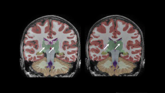
NeuroQuant® and its module, NeuroQuant MS, are leading medical device applications for volumetric MRI post-processing, providing automatic image segmentation and visualization of 75 brain structures, lesion counts, lesion burden and regional patterns. The software provides a slice-by-slice review of segmentation results, and disease-targeted reports to aid in the assessment of neurological conditions, disease progression and treatment effectiveness.
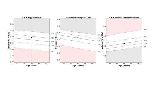
A distinguishing feature of NeuroQuant is its extensive normative database, the largest and most diverse available for clinical use, which allows NeuroQuant to compare each individual’s brain structure volumes to the general population. NeuroQuant automatically tracks brain structure and lesion volume changes over time, plotting previous exams as well as the current on normative reference charts, allowing at-a-glance assessment of disease progression.
NeuroQuant MS for the Assessment of Multiple Sclerosis
An integral component of multiple sclerosis (MS) management is accurate assessment of disease progression based on new and enlarging T2 FLAIR lesions. While MRI is the universally accepted tool for both diagnosis and monitoring of MS, manual lesion counts and comparisons are overly time-consuming in an increasingly RVU-focused world and inter-reader variability also poses challenges. NeuroQuant’s fully-automated analysis of MS lesion counts, volumes and change over time provides objective, reproducible results that correlate well with expert manual analyses1.
NeuroQuant MS also assists physicians in clinical evaluation, treatment planning and monitoring of disease progression by providing brain structure volumes in addition to lesion burden and dynamics. Fully-automated segmentation, measurement and charting of white and gray matter ROI volumes provide important diagnostic and prognostic information, particularly as the understanding of the relationship between gray matter volume loss and motor function deterioration increases. Automatic charting of structure volume changes over time also assists referrers in evaluating MS progression and the efficacy of disease-modifying therapies.
The FLAIR Lesion and Atrophy Report provides radiologists and referring physicians volumes of relevant brain structures as well as lesion counts, volumes and overall burden. Lesion reporting is provided by region, following McDonald criteria, and also hemispherically and by lobe. Brain structure volumes are compared to age- and sex-matched normative values while lesion dynamics information includes changes in counts and volumes of new, enlarging and shrinking lesions compared to that patient’s prior exam.
Lesion detection threshold settings are configurable to meet clinical needs. Minimum lesion size, separation, and detection sensitivity are customizable individually for the juxtacortical, periventricular, infratentorial, and white matter regions. Several color-coded lesion and brain structure reformats are available, including highly sensitive subtraction heat maps highlighting volume change in structures and pre-existing lesions. Outputs are automatically forwarded to the patient’s exam in PACS, typically in under 30 minutes.
Dr. Bash Reviews the NeuroQuant MS Quantitative Imaging Report for Multiple Sclerosis from Cortechs.ai on Vimeo.
Improving Radiology Throughput
Both NeuroQuant and NeuroQuant MS have been validated to work with SubtleMR, providing up to 60% reduction in scan time with the possibility of also increasing resolution and signal-to-noise ratio while denoising the scans. NeuroQuant is also compatible with OEM vendor-supported accelerated acquisitions and parallel imaging on 1.5 and 3T scanners.
References
Share