By
Cortechs.ai
2 mins
Amyloid-related imaging abnormalities (ARIA) are an assortment of MR imaging features identified in patients with Alzheimer’s disease treated with a new amyloid lowering drug regimen. They are common, dose-dependent effects of amyloid-targeting antibodies, strongly associated with the apolipoprotein E (APOE) ε4 allele1. ARIA is divided into two subtypes: vasogenic edema or sulcal effusion (ARIA-E) and cerebral microhemorrhages or superficial siderosis (ARIA-H), although they frequently co-occur2.
ARIA-E refers to the MR signal changes believed to represent vasogenic edema and related extravasated fluid. ARIA-E is hyperintense on FLAIR and T2W sequences and is typically transient. It is not associated with restricted diffusion or necrosis. At a cellular level, AIRA-E is believed to represent an increase in extracellular fluid volume due to the increase in brain capillary endothelial cell’s permeability to serum proteins3.
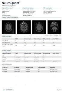
Cortechs.ai’s NeuroQuant® ARIA-E report uses conventional 2D or 3D FLAIR sequences to automatically quantify vasogenic edema in under 10 minutes. The report provides the volume, location, and count of regions of FLAIR hyperintensity, and offers tracking of new, enlarging, shrinking and stable edema when multi-time point MR scans are available.
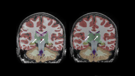
ARIA-H refers to microhemorrhages and superficial siderosis4. Microhemorrhages are small deposits of iron in tissue in the form of hemosiderin and are thought to represent the residue from small leakages of blood from a vessel into adjacent tissue parenchyma5. Microhemorrhages usually manifest as a focal, round, low intensity (relative to adjacent brain) lesions in the brain parenchyma, detected on T2* (GRE) weighted MRI sequences. Additional susceptibility weighting imaging (SWI) can be used to post-process and to improve microhemorrhage visualization6.
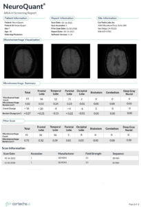
Cortechs.ai’s NeuroQuant ARIA-H report uses an additional 2D or 3D SWI or GRE (preferred) sequence to automatically quantify microhemorrhages in under 10 minutes. The report provides the count and location of microhemorrhage throughout the brain. When multi-time point MR scans are available, the report will display the interval change in the number and location of microhemorrhages and superficial siderosis.
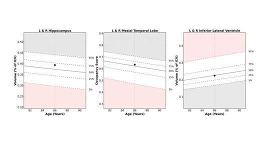
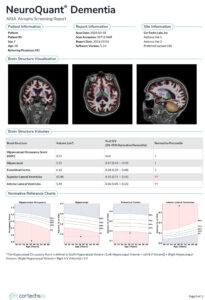
The ARIA screening report also includes a third page which presents the volumetric data of pertinent brain substructures that are important in dementia, such as the volume of the hippocampi, entorhinal cortex, and inferior lateral ventricles. The volumetric data is compared to a large normative age- and sex-matched database to determine the statistical significance of the degree of atrophy. These reports also allow for volumetric tracking over time when multi-time point studies are provided.
Cortechs.ai has developed the ARIA screening report to better characterize ARIA, quantify its occurrence, assess for rate of change over time, and to assist in clinical decision-making and treatment planning. The goal of the ARIA screening report is to aid neuroradiologists and neurologists by optimizing workflow, improving diagnostic accuracy, reducing reader subjectivity, and improving patient care.
References
Share